RPE lipofuscin is a yellow-orange pigment that builds up in the retinal pigment epithelium with age.
It forms from the daily turnover of photoreceptor outer segments.
Therefore, it is a marker of retinal aging and a driver of several diseases.
What RPE Lipofuscin Is
Lipofuscin is a mixture of fluorescent granules inside lysosomes of the pigment epithelium.
It cannot be broken down or removed from the cell.
Also, it accumulates throughout life.
As a result, the amount rises with age.
Origin from Outer Segments
Photoreceptors shed their outer segment tips each day.
The pigment epithelium phagocytoses these discs.
Also, retinoid byproducts of the visual cycle escape digestion.
Therefore, lipofuscin is a byproduct of vision itself.
Fundus Explorer Pro
Photograph the retinal findings described here with the phone already in your pocket — 22 D optics and built-in illumination in one handheld unit.
From Choroida — the team behind this siteBisretinoids and A2E
Sparrow and Boulton described the chemistry of RPE lipofuscin.
The main fluorophores are bisretinoids such as A2E.
Also, they form when vitamin A aldehyde reacts with phosphatidylethanolamine.
As a result, vitamin A metabolism links to the pigment.
Why RPE Lipofuscin Is Harmful
Bisretinoids generate reactive oxygen species when exposed to blue light.
They also act as detergents that damage membranes.
Also, they impair lysosomal function.
Therefore, high levels stress and kill the epithelial cell.
Fundus Autofluorescence
Delori and colleagues showed that the autofluorescence of the fundus comes mostly from RPE lipofuscin.
Blue light at 488 nanometers excites it.
Also, a confocal laser scanning device records the emission.
As a result, the image maps lipofuscin across the retina.
Normal Autofluorescence Pattern
The signal is dark at the fovea because of macular pigment.
It is brightest around the posterior pole and fades toward the periphery.
Also, the optic disc and vessels appear dark.
Therefore, a normal image has a typical pattern.
Increased Autofluorescence
Excess signal marks cells overloaded with lipofuscin.
Examples include vitelliform material and the edge of geographic atrophy.
Also, active lesions may show a bright halo.
As a result, high signal often marks disease activity.
Decreased Autofluorescence
Loss of RPE leads to a dark area.
This occurs in geographic atrophy and RPE tears.
Also, blood and thick media block the signal.
In addition, macular pigment darkens the fovea.
RPE Lipofuscin in Stargardt Disease
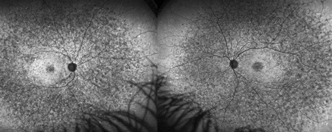
ABCA4 mutations impair clearance of retinoids from photoreceptors.
This causes massive buildup of bisretinoids in the epithelium (see Stargardt disease).
Also, the choroid appears dark on fluorescein angiography.
As a result, high lipofuscin explains the flecks and atrophy.
RPE Lipofuscin in Best Disease
Vitelliform material contains lipofuscin and outer segment debris.
The lesion is bright on autofluorescence.
Also, the signal falls as the material resolves.
Therefore, imaging tracks the stages of the disease.
RPE Lipofuscin in AMD
Holz and colleagues showed that patterns of increased autofluorescence at the border of atrophy predict its growth (see geographic atrophy).
Eyes with diffuse patterns progress faster.
Also, lipofuscin may contribute to cell death.
As a result, autofluorescence is a useful biomarker.
Therapies Targeting Lipofuscin
Visual cycle modulators aim to reduce bisretinoid formation.
Emixustat was tested in geographic atrophy and did not slow growth.
Also, Rosenfeld and colleagues reported these results in the SEATTLE study.
Therefore, the target remains under study.
Other Approaches to RPE Lipofuscin
Deuterated vitamin A slows dimer formation in models and is under trial in Stargardt disease.
Complement inhibitors target a different pathway in atrophy.
Also, antioxidants have not proved effective.
In addition, gene therapy may help in genetic forms.
Blue Light and Lipofuscin
Blue light exposure activates bisretinoids and may add stress.
The clinical effect of ordinary light is unclear.
Also, blue-blocking lenses lack proof of benefit.
Therefore, advise protection from intense light, not special glasses.
Using Autofluorescence in Practice
Use it to map atrophy and to follow progression.
It helps to distinguish flecks from drusen.
Also, it reveals early dystrophies before symptoms.
As a result, it belongs in the retinal work-up.
Limits of RPE Lipofuscin Imaging
Media opacity and image quality affect the signal.
Different devices give different brightness scales.
Also, interpretation needs experience.
Therefore, compare images from the same system.
Pitfalls to Avoid
- Do not read a dark fovea as atrophy
- Never compare images from different devices without care
- Do not assume that blue-blocking glasses prevent lipofuscin harm
- Never forget to check for pseudo-autofluorescence in poor-quality scans
Lipofuscin and Melanolipofuscin
In older eyes lipofuscin and melanin combine in granules called melanolipofuscin.
They reduce the protective role of melanin.
Also, they increase photosensitivity.
Therefore, aging changes the pigment mix.
For the latest studies, see the PubMed results for RPE lipofuscin.


Document what you see
Two smartphone imaging tools built for everyday clinic use — one for the slit lamp, one for the fundus.
From Choroida — the team behind this siteReferences
- Sparrow JR, Boulton M. RPE lipofuscin and its role in retinal pathobiology. Exp Eye Res. 2005;80:595-606.
- Delori FC, Dorey CK, Staurenghi G, Arend O, Goger DG, Weiter JJ. In vivo fluorescence of the ocular fundus exhibits retinal pigment epithelium lipofuscin characteristics. Invest Ophthalmol Vis Sci. 1995;36:718-729.
- Holz FG, Bindewald-Wittich A, Fleckenstein M, et al. Progression of geographic atrophy and impact of fundus autofluorescence patterns in age-related macular degeneration. Am J Ophthalmol. 2007;143:463-472.
- Rosenfeld PJ, Dugel PU, Holz FG, et al. Emixustat hydrochloride for geographic atrophy secondary to age-related macular degeneration: a randomized clinical trial. Ophthalmology. 2018;125:1556-1567.

Writes and reviews the clinical articles on Educate. Every post is checked against peer-reviewed sources before it goes live.