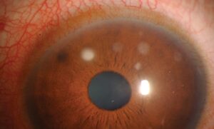
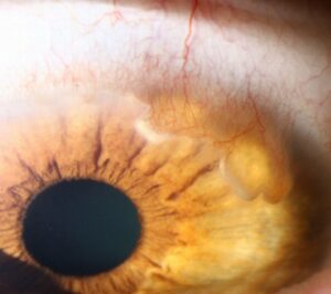
Racial melanosis is flat, benign conjunctival pigmentation seen in people with darker skin.
Clinicians also call it complexion-associated melanosis.
It is among the most common pigmented conjunctival findings in many clinics.
However, it can alarm patients because it resembles more serious lesions.
The clinician’s task is to recognize the pattern and notice when it breaks.
What Racial Melanosis Is
The finding reflects increased melanin within the basal conjunctival epithelium.
Melanocyte numbers may be normal or only mildly increased.
However, the cells show no atypia and no nested proliferation.
Therefore, it behaves as a stable physiological trait.
Who Develops It
It occurs mainly in people with darkly pigmented skin of many ancestries.
Pigment usually appears in childhood or early adulthood.
In addition, its extent tends to parallel skin complexion.
As a result, relatives often show the same finding.
All-fit Slit-Lamp Adapter
Record and share exactly what you see at the slit lamp. One adapter fits any slit lamp or surgical microscope — and any smartphone.
From Choroida — the team behind this siteClassic Features of Racial Melanosis
The pattern is the main diagnostic tool.
- Bilateral involvement, although often asymmetric
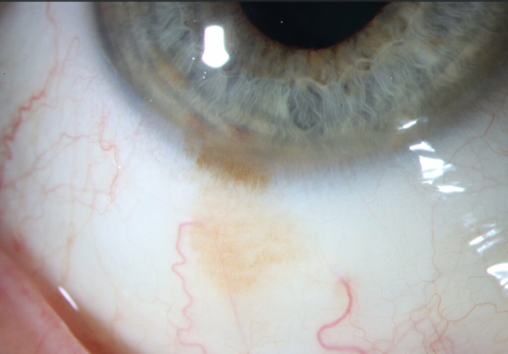
- Flat, brown, finely granular pigment
- Densest at the limbus, often as a perilimbal ring
- Interpalpebral bulbar conjunctiva most affected
- Pigment moves freely over the sclera
- No cysts, nodules or feeder vessels
Corneal Extension
Pigment may spread a short distance onto the peripheral cornea.
Sometimes it forms whorled streaks called striate melanokeratosis.
This pattern often follows epithelial injury or chronic surface disease.
Then, pigmented cells migrate centripetally with the healing epithelium.
Slit-Lamp Examination in Racial Melanosis
First, examine both eyes fully, including the fornices, tarsus and caruncle.
Next, slide the conjunctiva with a cotton tip to confirm epithelial pigment.
Also, use retroillumination to detect subtle elevation or vascular loops.
Finally, record clock hours and the estimated extent.
Imaging and Documentation
Serial slit-lamp photography provides the most useful baseline.
Anterior segment OCT shows a hyperreflective basal epithelial layer of normal thickness.
In contrast, melanoma often shows thickened tissue or an underlying mass.
Because imaging cannot replace histology, use it to support observation.
Racial Melanosis vs PAM
Primary acquired melanosis typically appears in middle-aged, fair-skinned adults.
It is usually unilateral and patchy, and it may wax and wane.
Also, it can extend into the fornix or palpebral conjunctiva.
PAM with atypia carries a real risk of melanoma (see primary acquired melanosis).
Therefore, new unilateral pigment in an adult deserves closer attention than lifelong bilateral pigment.
Racial Melanosis vs Conjunctival Melanoma
Melanoma is usually unilateral, elevated and vascular.
It may arise from PAM, a nevus, or de novo.
In addition, nonbulbar sites such as the fornix, tarsus and caruncle raise concern.
Conjunctival melanoma is uncommon in darker-skinned populations.
However, a nodule in a pigmented eye still needs biopsy, much as in the iris (see iris nevus versus melanoma).
Other Lookalikes
- Conjunctival nevus, often elevated with intralesional cysts
- Ocular melanocytosis, slate-gray scleral pigment that does not move
- Axenfeld nerve loop near the limbus
- Adrenochrome deposits from epinephrine drops
- Mascara deposits in the fornix
- Alkaptonuria with scleral pigment near muscle insertions
Red Flags in Racial Melanosis
Some changes break the benign pattern.
For example, new nodularity, growth on photos or feeder vessels demand action.
Also, pigment spreading into the fornix or tarsus needs evaluation.
Similarly, marked asymmetry developing in adulthood is suspicious.
When to Biopsy
Classic, stable, bilateral findings need no biopsy.
However, any red flag justifies biopsy by an ocular oncology team.
Mapping biopsies help when pigment is extensive or poorly defined.
Then, histology separates simple hyperpigmentation from PAM with atypia.
Follow-Up for Racial Melanosis
Typical cases need only routine eye examinations with photographs.
Compare each visit with the baseline image.
Meanwhile, ask patients to report new bumps, redness or rapid darkening.
As a result, you detect change early without excessive testing.
Taking the History in Racial Melanosis
First, ask when the patient or relatives first noticed the pigment.
Also, ask about prior eye injury, chronic inflammation or long-term drop use.
Then, review old photographs, because lifelong pigment strongly favors a benign cause.
In addition, ask about skin melanoma or atypical moles in the family.
Ocular Associations and Pregnancy
Hormonal change can darken existing conjunctival pigment in some patients.
For example, pregnancy may make a stable lesion look more intense.
However, darkening alone does not prove malignancy, so compare it with baseline photographs.
Therefore, document the change and recheck after delivery.
Counseling the Patient
Many patients notice the pigment in a mirror and fear cancer.
Explain that the color resembles skin pigment and usually stays stable.
Also, reassure them that appearance alone requires no drops or surgery.
However, encourage regular checkups.
Pitfalls to Avoid
- Do not label unilateral adult-onset pigment as racial melanosis without review
- Never skip lid eversion and fornix examination
- Avoid using skin color alone to exclude melanoma
- Do not treat benign pigment with cryotherapy or topical chemotherapy
- Always compare with prior photographs
For the latest studies, see the PubMed results for racial melanosis.


Document what you see
Two smartphone imaging tools built for everyday clinic use — one for the slit lamp, one for the fundus.
From Choroida — the team behind this siteReferences
- Shields CL, Demirci H, Karatza E, Shields JA. Clinical survey of 1643 melanocytic and nonmelanocytic conjunctival tumors. Ophthalmology. 2004;111:1747-1754.
- Shields JA, Shields CL, Mashayekhi A, et al. Primary acquired melanosis of the conjunctiva: risks for progression to melanoma in 311 eyes. Ophthalmology. 2008;115:511-519.
- Folberg R, McLean IW, Zimmerman LE. Primary acquired melanosis of the conjunctiva. Hum Pathol. 1985;16:129-135.
- Oellers P, Karp CL. Management of pigmented conjunctival lesions. Ocul Surf. 2012;10:251-263.
- Singh AD, Campos OE, Rhatigan RM, et al. Conjunctival melanoma in the black population. Surv Ophthalmol. 1998;43:127-133.

Writes and reviews the clinical articles on Educate. Every post is checked against peer-reviewed sources before it goes live.