Disciform keratitis recurrence means that stromal edema with keratic precipitates returns after a controlled episode.
Most cases reflect immune inflammation directed at herpes simplex antigen, and they often follow steroid reduction.
However, a returning disc can have other causes, so careful examination should come before escalation.
Why Disciform Keratitis Recurrence Happens
Herpes simplex virus stays latent in the trigeminal ganglion and can reactivate throughout life.
In addition, viral antigen may persist in the cornea and continue to stimulate inflammation.
Steroids suppress this reaction, while the latent virus stays in place.
Therefore, inflammation may return when treatment falls below the patient’s individual threshold (see herpetic disciform keratitis).
Recognizing the Returning Disc
Patients usually report blur, halos, photophobia or a dull ache.
The examiner typically finds a round zone of stromal edema under an intact epithelium.
Fine keratic precipitates lie beneath the disc, and reduced corneal sensation supports a herpetic cause.
All-fit Slit-Lamp Adapter
Record and share exactly what you see at the slit lamp. One adapter fits any slit lamp or surgical microscope — and any smartphone.
From Choroida — the team behind this siteExamination Pearls
First, test corneal sensation with a cotton wisp before instilling anesthetic drops.
Then compare both eyes, because a focal reduction supports prior herpetic disease.
In addition, stain with fluorescein to exclude a dendrite or geographic ulcer.
Finally, check intraocular pressure and look for endothelial changes.
Triggers of Disciform Keratitis Recurrence
Flares often follow rapid steroid withdrawal or the end of antiviral prophylaxis.
Ocular surgery and systemic immunosuppression may also precede an episode.
However, the Herpetic Eye Disease Study (HEDS) found no confirmed link with stress, sunlight, contact lens wear or eye injury.
Therefore, avoid promising patients that lifestyle changes will prevent flares.
Active Inflammation or Old Scar
Haze alone does not prove active disease.
Active inflammation shows edema, new keratic precipitates, increased thickness or anterior chamber cells.
In contrast, an old scar has defined borders and looks stable across serial photographs.
Next, compare pachymetry and anterior segment OCT with earlier records.
Steroids in Disciform Keratitis Recurrence
The HEDS stromal keratitis trial compared topical prednisolone with placebo in patients also receiving trifluridine.
Steroids reduced persistent or progressive inflammation and shortened the disease course.
However, visual outcome at six months did not differ significantly between groups.
Therefore, steroids control the episode, but they cannot guarantee long-term protection.
Tapering Without Triggering a Flare
The HEDS protocol tapered prednisolone over ten weeks, and many patients need longer in practice.
Reduce the frequency stepwise, then examine the eye after each meaningful reduction.
If a flare appeared at a known dose, slow the taper before reaching that level again.
No trial defines one ideal schedule, so clinical response should guide each decision.
Antiviral Cover During Steroid Therapy
Steroids can favor epithelial viral disease, especially at higher doses.
Consequently, many cornea specialists prescribe an antiviral throughout the steroid course.
Oral agents also avoid the surface toxicity of prolonged topical antivirals.
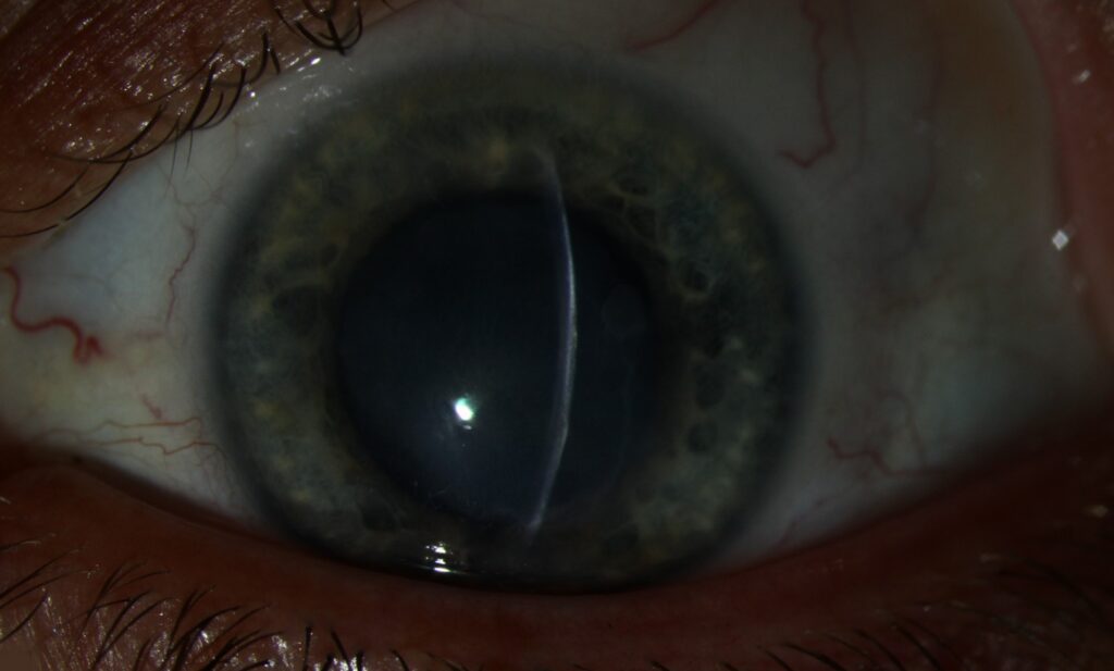
Preventing Disciform Keratitis Recurrence
The HEDS prophylaxis trial used oral acyclovir 400 mg twice daily for twelve months.
Ocular herpes recurred in 19% of treated patients and 32% of placebo patients.
Among patients with prior stromal keratitis, stromal recurrence occurred in 14% versus 28%.
Details of dosing and duration are discussed in acyclovir prophylaxis.
Limits of Long-Term Prophylaxis
Prophylaxis lowers risk, but breakthrough disease still occurs.
It also does not eradicate latent virus, so recurrences may follow discontinuation.
Therefore, weigh the number of episodes, visual potential, renal function and patient preference.
Mimics of Disciform Keratitis Recurrence
Consider these alternatives when the course or the examination looks atypical:
- Varicella zoster keratitis, especially with prior dermatomal rash or sectoral iris atrophy
- Cytomegalovirus endotheliitis, particularly with coin-shaped precipitates and raised pressure
- Corneal graft rejection, especially with an endothelial rejection line
- Acanthamoeba or fungal keratitis, particularly with severe pain or infiltrate
Poor response to appropriate treatment should also prompt aqueous PCR or corneal scrapings (see why keratitis treatment fails).
Steroid Complications During Long Courses
Long courses can raise intraocular pressure, so measure it at each visit.
In addition, prolonged treatment can promote posterior subcapsular opacity (see steroid-induced cataract).
Follow-Up After Disciform Keratitis Recurrence
Review the patient within one to two weeks after restarting treatment, then at longer intervals as edema resolves.
Also record pachymetry and visual acuity at each visit.
As a result, subtle relapse during the taper becomes easier to detect.
However, ask about symptoms between visits because early flares can be mild.
Counseling Patients
Explain that treatment controls inflammation and lowers risk, while it cannot remove the virus.
Next, warn patients never to stop steroids abruptly or restart old bottles without review.
Finally, ask them to report new redness, pain, halos or blur promptly.
Pitfalls to Avoid
- Do not start steroids before excluding an epithelial defect or infiltrate
- Never treat stable scar as active disease
- Avoid abrupt tapering after repeated flares
- Do not overlook elevated pressure during long steroid courses
For the latest studies, see the PubMed results for disciform keratitis recurrence.


Document what you see
Two smartphone imaging tools built for everyday clinic use — one for the slit lamp, one for the fundus.
From Choroida — the team behind this siteReferences
- Wilhelmus KR, Gee L, Hauck WW, et al. Herpetic Eye Disease Study: a controlled trial of topical corticosteroids for herpes simplex stromal keratitis. Ophthalmology. 1994;101:1883-1895.
- Herpetic Eye Disease Study Group. Acyclovir for the prevention of recurrent herpes simplex virus eye disease. N Engl J Med. 1998;339:300-306.
- Herpetic Eye Disease Study Group. Psychological stress and other potential triggers for recurrences of herpes simplex virus eye infections. Arch Ophthalmol. 2000;118:1617-1625.
- Herpetic Eye Disease Study Group. Predictors of recurrent herpes simplex virus keratitis. Cornea. 2001;20:123-128.
- White ML, Chodosh J. Herpes simplex virus keratitis: a treatment guideline. American Academy of Ophthalmology. 2014.

Writes and reviews the clinical articles on Educate. Every post is checked against peer-reviewed sources before it goes live.