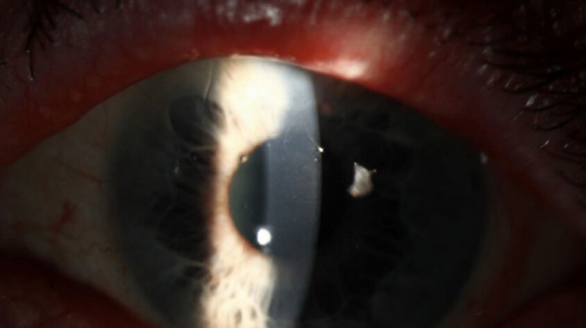
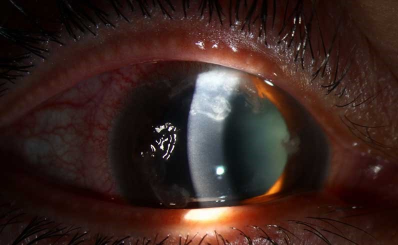
Satellite lesions are small, separate stromal infiltrates that surround a main corneal ulcer.
Clinicians classically link them with filamentous fungal keratitis, especially after agricultural trauma.
However, the sign only raises suspicion, and several other infections can reproduce it.
What Satellite Lesions Look Like
Each lesion appears as a small gray-white stromal opacity near the primary infiltrate.
Clear or mildly hazy cornea separates the two.
Also, the lesions may sit at different depths, so a single slit beam can miss them.
Examining Satellite Lesions at the Slit Lamp
First, survey the whole cornea with diffuse light, then narrow the beam through each opacity.
Next, use retroillumination, because faint foci often stand out against the red reflex.
Finally, record their positions, because a baseline map makes later comparison more reliable.
All-fit Slit-Lamp Adapter
Record and share exactly what you see at the slit lamp. One adapter fits any slit lamp or surgical microscope — and any smartphone.
From Choroida — the team behind this siteWhy Satellite Lesions Form
Fungal hyphae can extend through the stroma well beyond the visible infiltrate.
As a result, isolated foci may appear while the intervening cornea still looks clear.
Therefore, the visible ulcer often underestimates the true extent of infection.
History and Signs That Raise Suspicion
Trauma with plant matter is the classic history for filamentous fungal keratitis.
Contact lens wear and prior topical steroids also raise concern.
Serrated margins and a dry, raised slough point toward a filamentous organism.
Hypopyon occurs in both fungal and bacterial disease (see fungal versus bacterial clues).
Diagnostic Value of Satellite Lesions
Thomas and colleagues found that serrated margins, raised slough and non-yellow color helped predict filamentous infection.
Dalmon and colleagues showed that cornea specialists judging photographs classified ulcers correctly about two thirds of the time.
As a result, no single sign can replace microbiology.
Also, the absence of satellites does not exclude a fungal ulcer.
Mimics of Satellite Lesions
Nocardia can produce patchy pinhead infiltrates in a wreath pattern that resembles fungal disease (see Nocardia keratitis).
Nontuberculous mycobacteria also cause indolent ulcers with satellite foci, often after refractive surgery.
Acanthamoeba can cause multifocal infiltrates, so severe pain should prompt that diagnosis.
In addition, herpetic stromal keratitis may cause multifocal opacities, so check corneal sensation.
Confirming the Cause
Scrape the leading edge and ulcer base before starting therapy whenever possible (see stromal corneal ulcers).
KOH or calcofluor white can reveal hyphae quickly, while Gram stain screens for bacteria.
Then plate on blood, chocolate and Sabouraud media.
Finally, consider biopsy when deep infiltrates lie beneath intact epithelium.
Confocal Microscopy and Satellite Lesions
In vivo confocal microscopy can show branching hyphae within the stroma.
However, its yield depends on operator experience and lesion depth.
A negative scan does not exclude infection.
How Satellite Lesions Change Management
The sign should prompt same-day scraping and a lower threshold for antifungal treatment.
A positive smear justifies directed therapy before culture results return.
Central, deep or rapidly enlarging ulcers warrant urgent cornea review.
Antifungal Evidence
MUTT I found better outcomes with topical natamycin than with topical voriconazole, particularly in Fusarium ulcers.
MUTT II then found no significant reduction in perforation or therapeutic keratoplasty when oral voriconazole was added.
Therefore, topical natamycin is the usual starting point for filamentous disease.
However, its penetration is limited, and no drug can reverse established scarring.
Steroids and Satellite Lesions
Topical steroids can make an eye look quieter while fungi continue to multiply.
Therefore, withhold them while a fungal cause is still possible.
When Satellite Lesions Persist on Treatment
New or enlarging satellites after several days of therapy suggest resistance, poor penetration or a wrong diagnosis.
Therefore, repeat the scrape and review the culture and sensitivity results.
In addition, consider deeper sampling or biopsy if the stromal infiltrate keeps advancing.
Then discuss intrastromal or adjunctive options with a cornea colleague.
Follow-Up and Counseling
Record the epithelial defect, infiltrate size, satellites, hypopyon and thinning at each visit.
Explain that treatment often means frequent drops for weeks, because fungal ulcers heal slowly.
Also advise patients to stop contact lens wear and to protect the eye during outdoor work.
Finally, discuss scarring and possible transplantation early and honestly.
Pitfalls to Avoid
- Do not diagnose fungal keratitis from satellites alone
- Avoid ruling out fungus because satellites are absent
- Do not start steroids before excluding fungal infection
- Avoid delaying a repeat scrape when an ulcer worsens
For the latest studies, see the PubMed results for satellite lesions.


Document what you see
Two smartphone imaging tools built for everyday clinic use — one for the slit lamp, one for the fundus.
From Choroida — the team behind this siteReferences
- Thomas PA, Leck AK, Myatt M. Characteristic clinical features as an aid to the diagnosis of suppurative keratitis caused by filamentous fungi. Br J Ophthalmol. 2005;89:1554-1558.
- Dalmon C, Porco TC, Lietman TM, et al. The clinical differentiation of bacterial and fungal keratitis: a photographic survey. Invest Ophthalmol Vis Sci. 2012;53:1787-1791.
- Thomas PA, Kaliamurthy J. Mycotic keratitis: epidemiology, diagnosis and management. Clin Microbiol Infect. 2013;19:210-220.
- Prajna NV, Krishnan T, Mascarenhas J, et al. The Mycotic Ulcer Treatment Trial: a randomized trial comparing natamycin vs voriconazole. JAMA Ophthalmol. 2013;131:422-429.
- Prajna NV, Krishnan T, Rajaraman R, et al. Effect of oral voriconazole on fungal keratitis in the Mycotic Ulcer Treatment Trial II (MUTT II). JAMA Ophthalmol. 2016;134:1365-1372.

Writes and reviews the clinical articles on Educate. Every post is checked against peer-reviewed sources before it goes live.