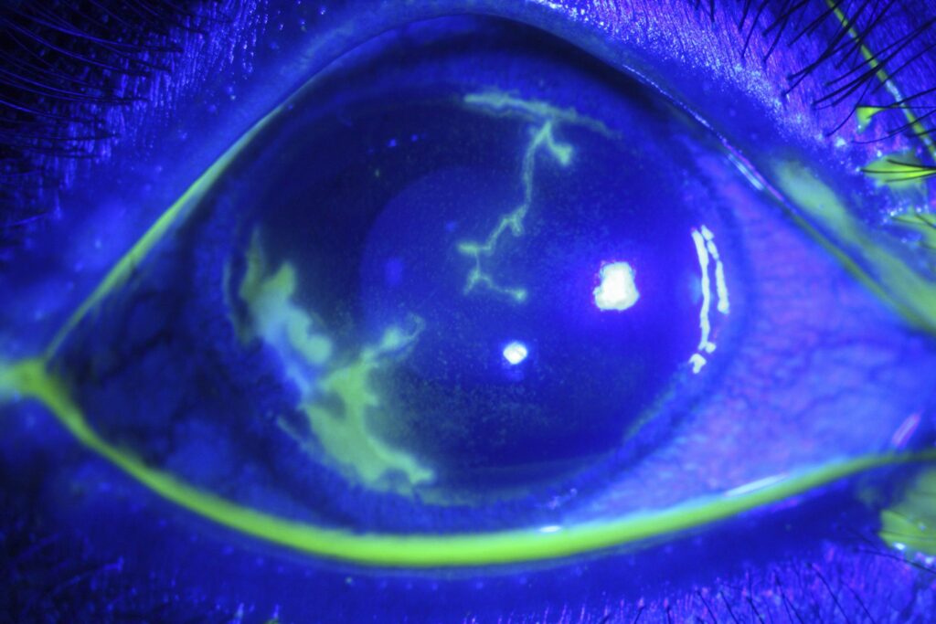
Dendritic ulcer is the classic corneal lesion of herpes simplex epithelial keratitis.
It appears as a branching epithelial defect with swollen ends called terminal bulbs.
Because the pattern is characteriztic, diagnosis usually rests on slit lamp findings.
However, mimics exist, and a wrong label can delay treatment or invite harmful steroids.
What a Dendritic Ulcer Is
The lesion reflects viral replication within corneal epithelial cells.
Most cases follow reactivation of latent virus from the trigeminal ganglion.
Therefore, many patients report earlier episodes, although a negative history does not exclude herpes.
What Patients Report
Patients describe redness, tearing, photophobia and foreign body sensation in one eye.
Blurring depends on whether the lesion crosses the visual axis.
However, pain may seem mild for the size of the defect because corneal sensation can fall.
Next, ask about cold sores and recent topical steroid use.
All-fit Slit-Lamp Adapter
Record and share exactly what you see at the slit lamp. One adapter fits any slit lamp or surgical microscope — and any smartphone.
From Choroida — the team behind this siteTerminal Bulbs in a Dendritic Ulcer
Terminal bulbs are the slightly expanded ends of each branch.
They help separate a herpetic dendrite from most pseudodendrites.
For example, a healing abrasion may branch, yet it usually lacks these bulbs.
Staining Pattern of a Dendritic Ulcer
Fluorescein pools in the ulcer bed because the epithelium is missing there.
In contrast, lissamine green stains the devitalized cells along the margins.
Atypical cases may need a broader review (see corneal staining patterns).
Corneal Sensation Testing
Test sensation before you instill any anesthetic.
Then compare both corneas with a cotton wisp.
Reduced sensation supports herpetic disease, although normal sensation does not exclude it.
Other Findings to Check
Conjunctival injection and a mild follicular reaction often accompany the epithelial lesion.
Next, inspect the lids for vesicles and measure intraocular pressure if the chamber is inflamed.
Finally, look beneath the ulcer for stromal haze or keratic precipitates, because deeper disease changes management.
Mimics of a Dendritic Ulcer
Several conditions can produce a branching epithelial figure:
- Herpes zoster pseudodendrites, which look raised and stain poorly (see herpes zoster ophthalmicus)
- Healing abrasions and recurrent erosions
- Acanthamoeba keratitis in a contact lens wearer with severe pain
- Toxic epitheliopathy from preservatives
- Tyrosinemia type II, which causes bilateral pseudodendrites in a child
When Testing Helps
Typical lesions rarely need laboratory confirmation.
However, consider PCR from a corneal swab when the course seems atypical.
Also, immunocompromised patients and children deserve a lower threshold for testing.
Contact Lens Wearers
Therefore, advise every lens wearer to stop wear until the epithelium heals.
Also, severe pain out of proportion to the findings should prompt corneal scraping to exclude Acanthamoeba.
However, do not delay antivirals when the lesion looks typical.
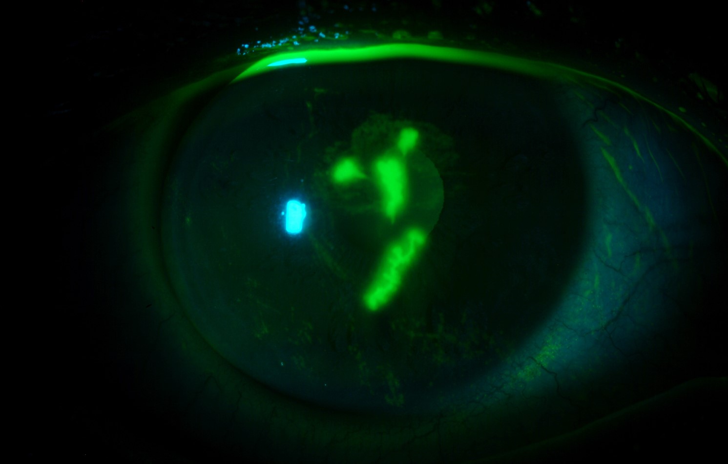
Topical Treatment of a Dendritic Ulcer
Topical antivirals inhibit viral DNA replication and speed healing.
Clinicians commonly give ganciclovir 0.15% gel five times daily until healing, then three times daily for seven days.
Trifluridine 1% is an alternative, usually given nine times daily.
However, prolonged trifluridine can injure the epithelium.
Cochrane evidence supports antivirals over placebo for epithelial keratitis.
Oral Antiviral Options
Oral therapy avoids surface toxicity.
Typical regimens include acyclovir 400 mg five times daily or valacyclovir 500 mg two to three times daily.
Courses usually last 7 to 10 days, and renal function may require dose adjustment.
Role of Debridement
Gentle debridement removes infected epithelium and reduces viral load.
However, it cannot prevent later recurrence.
Therefore, most clinicians combine it with antivirals.
Steroids and a Dendritic Ulcer
Topical steroids can enlarge an active lesion into a geographic ulcer.
Therefore, avoid them while infectious epithelial disease persists.
Stromal keratitis may later require steroids under antiviral cover (see herpes simplex keratitis).
Follow-Up of a Dendritic Ulcer
Review the patient within about a week.
Most ulcers heal within one to two weeks on appropriate therapy.
Persistent staining should prompt a review of adherence, drug toxicity and the diagnosis.
In addition, a smooth nonhealing defect may indicate a metaherpetic ulcer.
Recurrence Triggers
Fever, ultraviolet exposure, trauma and emotional stress may precipitate reactivation.
Also, ocular surgery and laser procedures can trigger recurrence in susceptible eyes.
Therefore, ask about recent triggers at each visit.
However, many recurrences occur without an obvious cause.
Preventing Recurrence
The Herpetic Eye Disease Study tested oral acyclovir 400 mg twice daily for 12 months.
Ocular recurrence fell to 19% with acyclovir, compared with 32% on placebo.
Therefore, reserve long-term suppression for frequent or visually threatening recurrences.
Counseling a Patient With a Dendritic Ulcer
Explain that treatment clears active disease, while the virus stays latent.
Consequently, recurrence remains possible.
Finally, ask patients to return early for new pain, redness or blur.
Pitfalls to Avoid
- Do not prescribe a steroid-antibiotic combination for an undiagnosed red eye
- Avoid diagnosing herpes from branching alone without checking for terminal bulbs
- Never continue topical antivirals indefinitely for persistent staining
- Always test sensation before anesthetic drops
For the latest studies, see the PubMed results for dendritic ulcer.


Document what you see
Two smartphone imaging tools built for everyday clinic use — one for the slit lamp, one for the fundus.
From Choroida — the team behind this siteReferences
- Herpetic Eye Disease Study Group. Acyclovir for the prevention of recurrent herpes simplex virus eye disease. N Engl J Med. 1998;339:300-306.
- Wilhelmus KR. Antiviral treatment and other therapeutic interventions for herpes simplex virus epithelial keratitis. Cochrane Database Syst Rev. 2015;1:CD002898.
- Austin A, Lietman T, Rose-Nussbaumer J. Update on the management of infectious keratitis. Ophthalmology. 2017;124:1678-1689.
- Liesegang TJ. Herpes simplex virus epidemiology and ocular importance. Cornea. 2001;20:1-13.

Writes and reviews the clinical articles on Educate. Every post is checked against peer-reviewed sources before it goes live.