Glaucoma screening aims to find a disease that causes no symptoms early.
Half of affected people do not know they have it.
Targeted case-finding is more useful than blanket tests.
Why Glaucoma Screening Is Difficult
Early open-angle glaucoma causes no pain and no visual change.
Pressure alone misses many cases.
No single test is both sensitive and specific.
Therefore, screening needs a combination of tests.
What the USPSTF Concluded
The US Preventive Services Task Force reviewed the evidence in 2022.
It found insufficient evidence to recommend population screening.
This reflects a lack of long-term trial data.
So the statement does not mean glaucoma is unimportant.
All-fit Slit-Lamp Adapter
Record and share exactly what you see at the slit lamp. One adapter fits any slit lamp or surgical microscope — and any smartphone.
From Choroida — the team behind this siteWho Needs Glaucoma Screening
Adults over 60 years have higher risk.
People of African ancestry develop disease earlier.
A family history raises the risk several-fold.
Also include patients with high myopia and diabetes.
Glaucoma Screening and Family History
First-degree relatives of patients have a higher risk.
Start examinations earlier in this group.
Repeat them at regular intervals.
Therefore, encourage patients to alert family members.
Intraocular Pressure Measurement
Applanation tonometry gives a reliable reading (see Goldmann applanation tonometry).
A normal pressure does not exclude glaucoma.
Many patients have disease at normal pressure (see normal-tension glaucoma).
So never rely on pressure alone.
Optic Nerve Examination
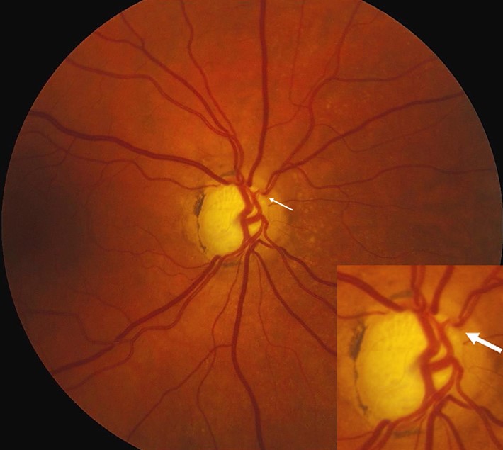
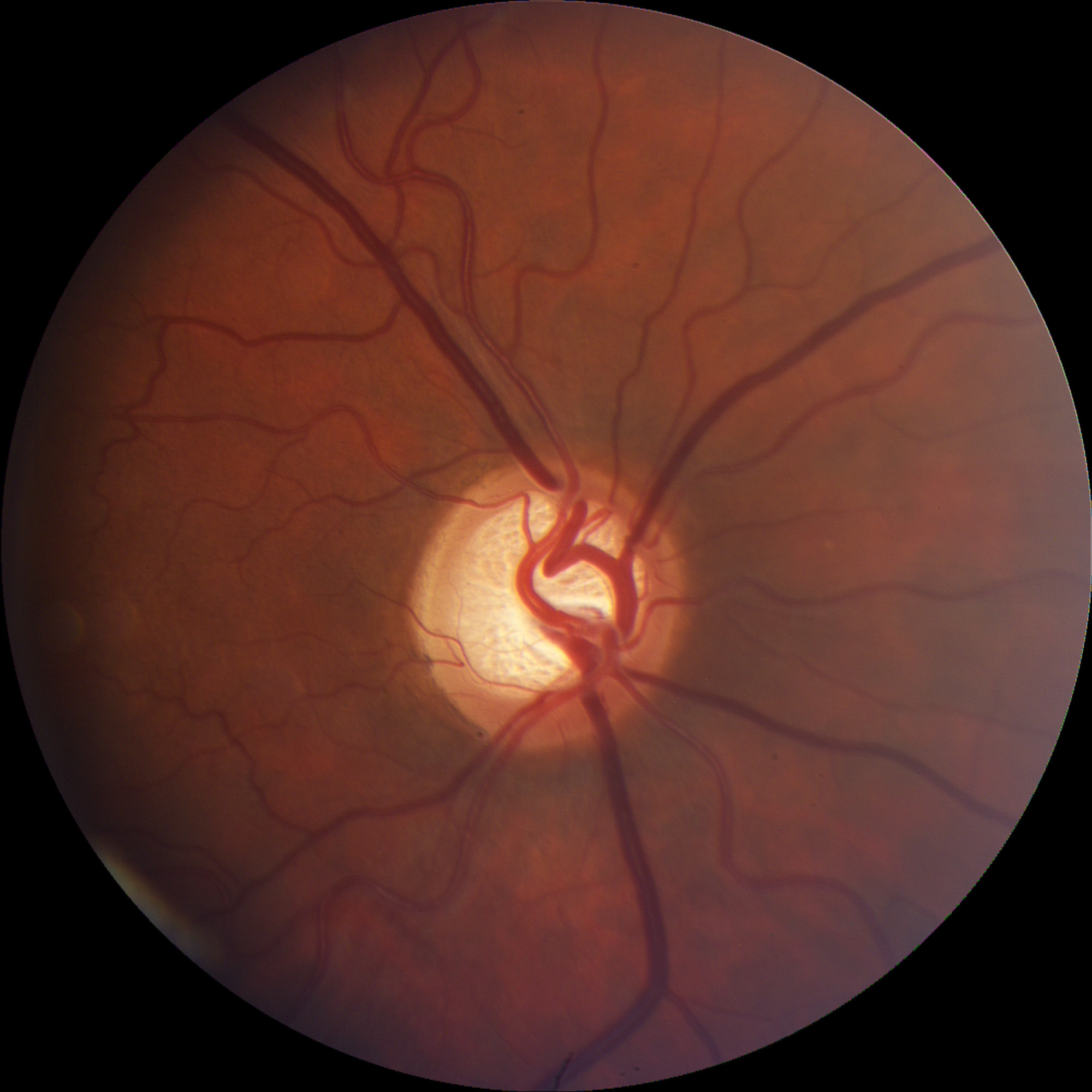
Look at the cup-to-disc ratio and rim shape.
Check for notches, hemorrhages and asymmetry.
Photographs allow comparison over time.
Therefore, examine the disc after dilation when possible.
Visual Field Testing for Glaucoma Screening
Automated perimetry detects functional loss.
Early defects are subtle.
Testing needs good cooperation.
So repeat abnormal results before making a diagnosis.
OCT in Glaucoma Screening
OCT measures retinal nerve fiber layer thickness.
It is useful for detecting early loss.
Artifacts and myopia can mislead.
Also interpret it with the clinical picture.
Gonioscopy in Glaucoma Screening
Angle closure is a second form that screening must catch.
Gonioscopy examines the angle directly.
Narrow angles need monitoring or laser treatment (see primary angle closure suspect).
Therefore, do not skip it.
Case-Finding in Clinics
Optometrists and ophthalmologists find most new cases during routine visits.
Review disc, pressure and angle in every adult over 40.
Refer suspicious findings early.
So opportunistic detection is effective.
Telemedicine and AI
Fundus photographs read by software can flag suspicious discs.
These tools expand access in remote areas.
They still need clinician review.
Also validate them in the local population.
Counseling About Glaucoma Screening
Explain that glaucoma is silent and treatable.
Encourage regular examinations for high-risk patients.
Give clear information about what to expect.
Therefore, patients understand the value of follow-up.
Pitfalls to Avoid
- Do not rely on pressure alone
- Never skip gonioscopy in suspects
- Avoid labeling from a single visual field
- Do not forget family history
Glaucoma Screening in Diabetes
Diabetes raises the risk of open-angle disease.
Because diabetic patients already have eye examinations, add pressure and disc review.
If both are normal, continue routine care.
Therefore, combine the two screening programs.
Glaucoma Screening and Race
People of African ancestry develop glaucoma earlier and more severely.
Because the risk starts around the age of 40, begin screening then.
If a relative is affected, start sooner.
Therefore, tailor the starting age to ancestry.
Glaucoma Screening in Asian Populations
Angle closure is more common in Asian populations.
When screening, assess the angle in addition to the disc.
Because dilation can trigger an attack, check the angle first.
Therefore, gonioscopy has extra value here.
Glaucoma Screening Costs
Screening large groups is costly and finds many false positives.
Because targeted programs are more efficient, focus on high-risk groups.
If resources are limited, prioritize relatives of patients.
Then invest in follow-up for confirmed cases.
Glaucoma Screening Frequency
Adults at average risk may need an examination every few years.
High-risk adults need annual or biennial checks.
Because disease can start at any time, repeat the assessment.
Then adjust the interval to the findings.
Glaucoma Screening and Referral
Refer patients with suspicious discs, high pressure or narrow angles.
Because delays harm vision, use clear pathways.
If testing is borderline, arrange repeat visits.
Therefore, communicate with the referring practitioner.
Glaucoma Screening: Practical Summary
Test adults at higher risk, such as those with a family history or African ancestry.
Because pressure alone misses disease, combine it with disc and field assessment.
If the angle is narrow, arrange further tests.
Finally, make sure every positive result reaches a specialist.
For the latest studies, see the PubMed results for glaucoma screening.


Document what you see
Two smartphone imaging tools built for everyday clinic use — one for the slit lamp, one for the fundus.
From Choroida — the team behind this siteReferences
- Owens DK, Davidson KW, Krist AH, et al. Screening for primary open-angle glaucoma: US Preventive Services Task Force recommendation statement. JAMA. 2022;327:1992-1997.
- Tham YC, Li X, Wong TY, Quigley HA, Aung T, Cheng CY. Global prevalence of glaucoma and projections of glaucoma burden through 2040. Ophthalmology. 2014;121:2081-2090.
- Gedde SJ, Vinod K, Wright MM, et al. Primary open-angle glaucoma preferred practice pattern. Ophthalmology. 2021;128:P71-P150.
- Weinreb RN, Aung T, Medeiros FA. The pathophysiology and treatment of glaucoma: a review. JAMA. 2014;311:1901-1911.

Writes and reviews the clinical articles on Educate. Every post is checked against peer-reviewed sources before it goes live.