
Primary angle closure suspect (PACS) describes an eye with an anatomically narrow angle, in which the trabecular meshwork is hidden by the iris, but the intraocular pressure and the optic disc are normal.
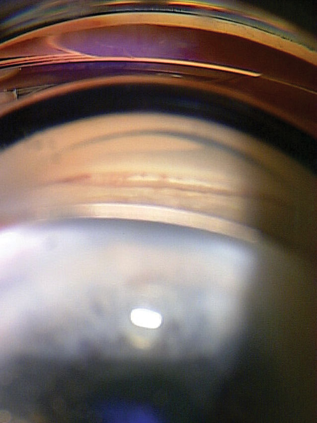
It is the earliest stage of the primary angle closure spectrum, and it is common in some populations.
Most eyes with PACS never develop glaucoma, and the clinical challenge is to identify the minority at risk.
Primary Angle Closure Suspect and the Angle Closure Spectrum
The International Society of Geographical and Epidemiological Ophthalmology classification divides the spectrum into three stages.
- PACS: iridotrabecular contact in at least 180 degrees on gonioscopy, normal pressure, no peripheral anterior synechiae, and a normal optic nerve
- Primary angle closure (PAC): PACS with raised pressure or peripheral anterior synechiae, but no glaucomatous optic neuropathy
- Primary angle closure glaucoma (PACG): PAC with glaucomatous optic neuropathy
The distinction matters because each stage carries a different risk and may call for a different treatment.
Risk Factors
Angle closure occurs more often in older patients, women, hyperopes, and people of East Asian or Inuit ancestry.
A positive family history increases the risk, and the biometric features include a short axial length, a shallow anterior chamber, a thick anterior lens, and a forward lens position.
Some medications, such as anticholinergics and topiramate, can precipitate closure in a susceptible eye.
Pupillary block is the most common mechanism, and a plateau iris configuration can contribute (see plateau iris).
All-fit Slit-Lamp Adapter
Record and share exactly what you see at the slit lamp. One adapter fits any slit lamp or surgical microscope — and any smartphone.
From Choroida — the team behind this siteExamination
Gonioscopy is the key examination, and it should be done in dim light with a small beam that does not constrict the pupil.
Use a static view without indentation first, and then a dynamic view with a four-mirror lens to open an appositional angle.
The finding to look for is how much of the posterior trabecular meshwork can be seen, and whether there are synechiae.
Anterior segment OCT is a useful adjunct that provides an objective and repeatable assessment of angle width (see anterior segment OCT).
Measure the pressure, examine the optic disc, and perform visual fields and OCT of the nerve fiber layer to establish a baseline.
Does Iridotomy Help?
The Zhongshan Angle Closure Prevention trial randomized people with PACS to laser peripheral iridotomy in one eye and observation in the other.
Over six years, the treated eye had a lower rate of progression to PAC, and the risk reduction was about half, although the absolute incidence was low in both eyes.
This suggests that the benefit is real but modest, and that many patients will not progress even without treatment.
Because of this, practice varies, and the decision depends on the degree of angle narrowing, the family history, the ability of the patient to follow up, and the access to emergency care.
Management
Offer iridotomy to patients at higher risk, including those with a strong family history, with very narrow angles, who live far from care, or who need a medication that might precipitate closure (see laser peripheral iridotomy).
Other patients can be observed with regular gonioscopy, pressure checks, and optic nerve assessment.
Lens extraction has a role in older patients with PAC or PACG, and the results of recent trials support it in this group.
Whatever the choice, all patients should learn the warning symptoms of an acute attack: eye pain, blur, halos, and headache (see acute angle closure).
Provocative Testing and Imaging
Provocative tests such as the dark room test have limited predictive value, and they are not widely used.
Anterior segment OCT and ultrasound biomicroscopy show the iris configuration, the lens vault, and the anterior chamber depth, and they can show whether the angle is closed because of pupillary block, plateau iris, or a thick lens.
These images help to choose between iridotomy and lens extraction, and they provide a baseline for follow-up.
The most helpful feature on OCT is often the lens vault, which is the height of the anterior lens surface above the line joining the two scleral spurs.
Counselling the Patient
Explain that a narrow angle is an anatomical feature, that it does not mean the person has glaucoma, and that most people with it never lose vision.
Describe the symptoms of an acute attack, and give written advice about when to seek urgent care.
Mention that certain medicines, such as some antihistamines, antidepressants, and decongestants, can dilate the pupil and may need caution, although the risk is low in most patients.
Follow-Up
After iridotomy, repeat gonioscopy to confirm that the angle has opened, and measure the pressure.
Patients managed by observation need examination at least yearly, with more frequent visits when the angle is very narrow or when changes appear.
The development of synechiae or a rise in pressure changes the classification and the management.


Document what you see
Two smartphone imaging tools built for everyday clinic use — one for the slit lamp, one for the fundus.
From Choroida — the team behind this siteReferences
- Foster PJ, Buhrmann R, Quigley HA, Johnson GJ. The definition and classification of glaucoma in prevalence surveys. Br J Ophthalmol. 2002;86:238-242.
- He M, Jiang Y, Huang S, et al. Laser peripheral iridotomy for the prevention of angle closure: a single-centre, randomised controlled trial. Lancet. 2019;393:1609-1618.
- Azuara-Blanco A, Burr J, Ramsay C, et al. Effectiveness of early lens extraction for the treatment of primary angle-closure glaucoma (EAGLE). Lancet. 2016;388:1389-1397.
- American Academy of Ophthalmology. Basic and Clinical Science Course, Section 10: Glaucoma. AAO; current edition.

Writes and reviews the clinical articles on Educate. Every post is checked against peer-reviewed sources before it goes live.


