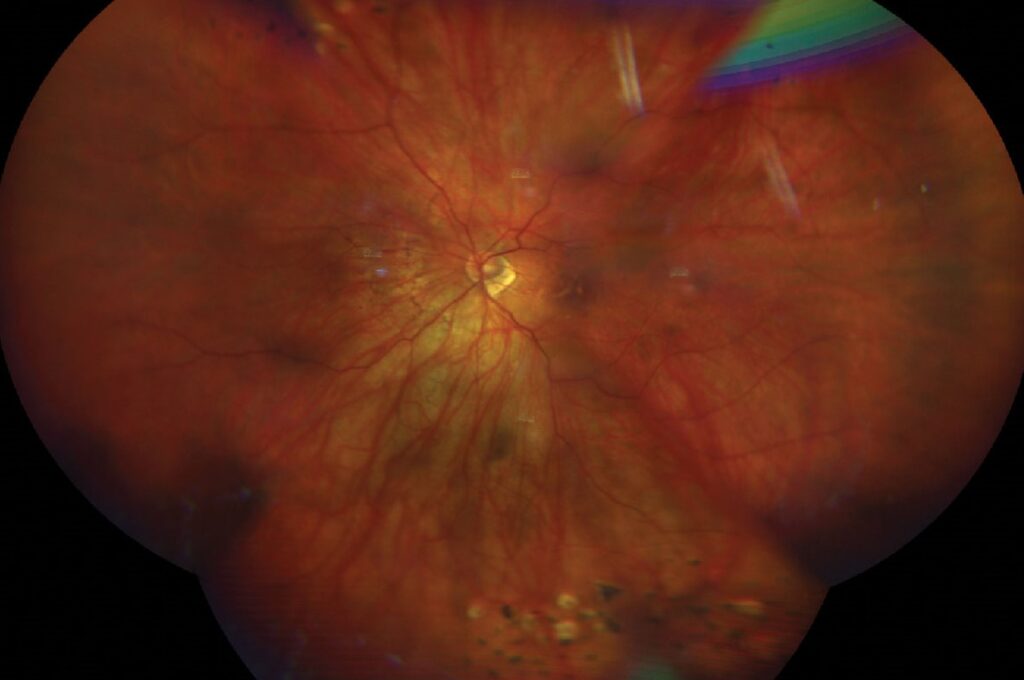
A tessellated fundus shows the choroidal vessels clearly through the retina.
The orange-red polygons look like a mosaic between darker vessels.
Therefore, the sign reflects thin pigment, thin choroid or both.
What a Tessellated Fundus Looks Like
The fundus background shows a pattern of large choroidal vessels separated by pigmented islands.
The pattern is clearest near the posterior pole and the disc.
Also, it is seen in light-skinned people and in many myopic eyes.
As a result, it is one of the commonest fundus findings.
Why a Tessellated Fundus Appears
The retinal pigment epithelium normally masks the choroid.
When the pigment thins or the choroid shrinks, the vessels show through.
Also, a thin retina adds to transparency.
Therefore, several mechanisms can create the same appearance.
Fundus Explorer Pro
Photograph the retinal findings described here with the phone already in your pocket — 22 D optics and built-in illumination in one handheld unit.
From Choroida — the team behind this siteTessellated Fundus in Normal Eyes
Fair-skinned or blond people can show the pattern without disease.
Older age increases the chance.
Also, many young myopic adults show mild tessellation.
As a result, the finding alone does not prove disease.
Link with Myopia
Axial elongation stretches the retina and thins the choroid.
Tessellation rises with axial length and myopic refraction.
Also, Yan and colleagues found tessellation in a large share of adults in the Beijing Eye Study.
Therefore, it is an early sign of structural change.
Tessellated Fundus in the META-PM Classification
The META-PM system grades myopic maculopathy from none to macular atrophy.
Category one is a tessellated fundus only.
Also, higher categories include diffuse and patchy atrophy (see META-PM classification).
As a result, tessellation is the starting point of the scale.
Choroidal Thickness
Tessellation correlates with thin subfoveal choroid on OCT.
The choroid is thinner in eyes with higher density of the sign.
Also, choroidal thinning may precede the visible change.
Therefore, OCT can quantify what the eye shows.
Fundus Tessellation Density
Researchers measure the density of the sign by image analysis of the fundus photograph.
The density gives a numeric measure that tracks myopia progression.
Also, the measure correlates with choroidal vessel visibility.
In addition, it can help in large studies of children.
Tessellation in Children
Children with progressive myopia may show tessellation early.
It signals choroidal thinning even when the refraction is modest.
Also, myopia control treatment aims to slow this process.
Therefore, document the finding at baseline.
Other Causes of a Tessellated Fundus
Albinism and oculocutaneous hypopigmentation show tessellation through lack of pigment.
Choroidal sclerosis in older adults can show a similar pattern.
Also, some inherited dystrophies thin the pigment layer.
As a result, review the history and the other findings.
When a Tessellated Fundus Signals Disease
Patchy atrophy, lacquer cracks or staphyloma suggest advanced myopic disease.
These changes raise the risk of vision loss.
Also, a change in the pattern over time deserves attention.
Therefore, look at the whole fundus and not at tessellation alone.
Differentiating from Choroidal Atrophy
Atrophy shows bare sclera and sharply outlined pale areas.
Tessellation shows intact pigment islands between vessels.
Also, large vessels in atrophy appear white and stripped.
In addition, OCT shows loss of the pigment epithelium in atrophy.
Follow-Up of a Tessellated Fundus
In high myopia, examine the fundus yearly.
Use OCT of the macula to detect early tractional or atrophic changes (see retinal detachment in high myopia).
Also, advise patients about warning signs of detachment.
Therefore, regular review protects vision.
Imaging Options
Color photography records the pattern well.
Red-free and infrared images show the choroidal vessels more clearly.
Also, widefield imaging captures the peripheral pattern.
As a result, choose the mode that fits the question.
Counseling Patients
Explain that the pattern is common and often harmless.
Say that it can be a sign of a long eye and thin tissue.
Also, suggest yearly checks if myopia is high.
In addition, remind them to report flashes or shadows.
Pitfalls to Avoid
- Do not label every tessellated fundus as pathologic myopia
- Never forget to check the periphery for lattice and tears
- Do not confuse tessellation with choroidal atrophy
- Never skip follow-up in children with fast myopia progression
Choroidal Vessel Visibility and OCT
En face OCT of the choroid shows the large vessels that cause the pattern.
The vessel layer thins as tessellation increases.
Also, the scleral line may look more prominent.
Therefore, OCT confirms the clinical impression.
Myopia Control and the Fundus
Treatments such as atropine and orthokeratology aim to slow axial growth.
Slower growth may slow the choroidal thinning.
Also, evidence on fundus changes is still limited.
As a result, track both refraction and fundus.
For the latest studies, see the PubMed results for tessellated fundus.


Document what you see
Two smartphone imaging tools built for everyday clinic use — one for the slit lamp, one for the fundus.
From Choroida — the team behind this siteReferences
- Yan YN, Wang YX, Xu L, Xu J, Wei WB, Jonas JB. Fundus tessellation: prevalence and associated factors: the Beijing Eye Study 2011. Ophthalmology. 2015;122:1873-1880.
- Ohno-Matsui K, Kawasaki R, Jonas JB, et al. International photographic classification and grading system for myopic maculopathy. Am J Ophthalmol. 2015;159:877-883.
- Ohno-Matsui K, Wu PC, Yamashiro K, et al. IMI pathologic myopia. Invest Ophthalmol Vis Sci. 2021;62(5):5.
- Nickla DL, Wallman J. The multifunctional choroid. Prog Retin Eye Res. 2010;29:144-168.

Writes and reviews the clinical articles on Educate. Every post is checked against peer-reviewed sources before it goes live.