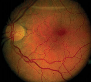
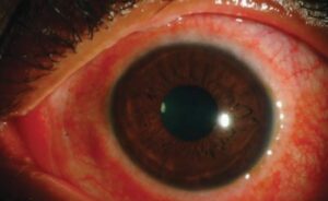
Anemic retinopathy is the set of retinal changes that appear in patients with severe anemia, often with low platelets.
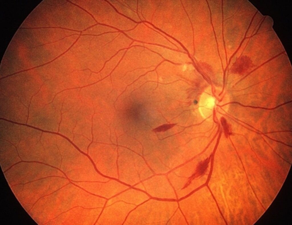
It includes flame and dot hemorrhages, cotton wool spots, Roth spots, and venous dilatation.
The findings usually reverse when the blood counts recover, but they may mark serious systemic disease.
What Anemic Retinopathy Is
Low hemoglobin reduces oxygen delivery to the retina, so retinal capillaries become leaky and fragile.
Thrombocytopenia adds a bleeding tendency, and the two together raise the risk of hemorrhage.
Rubenstein and colleagues described the combination of anemia, thrombocytopenia, and retinal hemorrhage in the 1960s.
Carraro and colleagues later found retinopathy in a substantial share of patients with severe anemia or low platelets (see Roth spots).
Signs of Anemic Retinopathy
- Flame-shaped and dot-blot hemorrhages in the posterior pole
- Roth spots, which are hemorrhages with a pale center
- Cotton wool spots from nerve fiber layer ischemia
- Dilated and tortuous veins, and a pale fundus
Preretinal and vitreous hemorrhage can also occur when platelet counts are very low (see retinal hemorrhage patterns).
Fundus Explorer Pro
Photograph the retinal findings described here with the phone already in your pocket — 22 D optics and built-in illumination in one handheld unit.
From Choroida — the team behind this siteCauses of Anemic Retinopathy
The condition occurs in iron deficiency, vitamin B12 deficiency, aplastic anemia, hemolytic anemia, and blood loss.
Leukemia and other marrow disorders cause similar changes through anemia, thrombocytopenia, and infiltration.
Pregnancy, malnutrition, and chronic kidney disease can also lead to anemia severe enough to affect the retina (see leukemic retinopathy).
Examination and Workup
Dilate the pupil and examine the whole fundus, since hemorrhages may lie outside the posterior pole.
Document the findings with photographs and OCT.
Order a complete blood count, a reticulocyte count, and tests for the cause of the anemia.
Refer promptly to a physician when blood counts are very low or when other signs of bleeding exist.
Differential Diagnosis of Anemic Retinopathy
- Diabetic and hypertensive retinopathy
- Retinal vein occlusion and ocular ischemic syndrome
- Leukemic retinopathy and infective endocarditis
- Valsalva and Purtscher retinopathy
The blood tests and the systemic history usually separate these (see hypertensive retinopathy).
Treatment and Outcome of Anemic Retinopathy
Treat the cause of the anemia and correct the low platelet count.
Transfusion or iron replacement raises hemoglobin, and the hemorrhages usually clear over weeks.
Vision recovers in most patients, although macular hemorrhage may leave a scar.
Rare vitreous hemorrhage may need observation or vitrectomy.
An Example Case
A 34-year-old woman with heavy menstrual bleeding reports blurred vision in both eyes for a week.
Her hemoglobin is 4.8 g/dL, and examination shows scattered flame hemorrhages, a few cotton wool spots, and one Roth spot in the right eye.
Her acuity is 20/30, and the macula is spared.
After transfusion and iron replacement, the hemorrhages fade over a month, and her vision returns to 20/20.
Questions Patients Ask
Patients ask whether the eye damage is permanent, and the answer is that it usually clears when the blood count recovers.
They also ask whether they need eye surgery, and they rarely do.
Because the eye findings reflect a body-wide problem, explain the role of the medical team.
Pitfalls to Avoid
- Treating the retina as diabetic or hypertensive without checking the blood count
- Missing leukemia in a patient with hemorrhages and low platelets
- Forgetting to repeat the examination after treatment
- Overlooking the macula, where a hemorrhage may affect vision
Looking Ahead
Retinal photographs may help to track the response to treatment in resource-limited settings.
Also, simple screening tools may detect severe anemia from retinal signs, though evidence is limited.
Therefore, the eye exam may support, but not replace, laboratory testing.
Practical Points
Order a blood count the same day when the fundus shows bilateral hemorrhages without an obvious cause.
Then arrange follow-up at two to four weeks to confirm resolution.
Also record photographs, so that later comparison is easy.
Anemia and the Eye in Other Forms
Severe anemia may also cause optic neuropathy, conjunctival pallor, and subconjunctival hemorrhage.
Sickle cell disease and thalassemia produce their own retinal changes.
Therefore, always consider the type of anemia when you interpret the fundus.
Systemic Evaluation
Ask about bleeding, diet, medications, and menstrual history.
Then examine the patient for pallor, petechiae, and enlarged lymph nodes.
Because severe anemia may signal leukemia, order a peripheral smear.
Also check platelets and clotting studies when hemorrhages are present.
Follow-Up
Re-examine the retina after the blood count improves.
Then confirm that the hemorrhages resolve.
Also document visual acuity at each visit.
Warning Signs for Patients
Teach patients to return if they notice new floaters or sudden loss of vision.
Also ask them to report ongoing bleeding or fatigue to their physician.
Then arrange a prompt review when symptoms change.


Document what you see
Two smartphone imaging tools built for everyday clinic use — one for the slit lamp, one for the fundus.
From Choroida — the team behind this siteReferences
- Rubenstein RA, Yanoff M, Albert DM. Thrombocytopenia, anemia, and retinal hemorrhage. Am J Ophthalmol. 1968;65:435-439.
- Carraro MC, Rossetti L, Gerli GC. Prevalence of retinopathy in patients with anemia or thrombocytopenia. Eur J Haematol. 2001;67:238-244.
- Holt JM, Gordon-Smith EC. Retinal abnormalities in diseases of the blood. Br J Ophthalmol. 1969;53:145-160.
- American Academy of Ophthalmology. Basic and Clinical Science Course, Section 12: Retina and Vitreous. AAO; current edition.

Writes and reviews the clinical articles on Educate. Every post is checked against peer-reviewed sources before it goes live.