
Rickettsial retinitis is a retinal inflammation that occurs in patients infected with Rickettsia species, which are transmitted by ticks, fleas, lice, or mites.
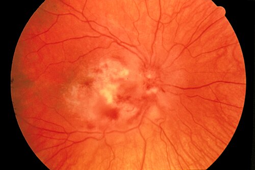
It is a treatable cause of infectious retinal vasculitis, and the clue is a febrile illness with a rash in a patient who has retinal lesions.
Most patients recover well when treated with doxycycline, so recognition matters.
What Is Rickettsial Retinitis?
Rickettsiae are intracellular bacteria that infect the endothelial cells of small vessels, causing vasculitis.
In the eye, this leads to retinal vessel inflammation, hemorrhage, and ischemia.
The species most often reported include Rickettsia conorii, the cause of Mediterranean spotted fever, Rickettsia typhi, which causes murine typhus, and Rickettsia rickettsii, the cause of Rocky Mountain spotted fever.
Scrub typhus, caused by a related bacterium, can produce similar retinal findings.
Systemic Features
The illness starts with fever, headache, and muscle pain.
A maculopapular or petechial rash appears a few days later, and it may involve the palms and soles.
An eschar at the tick bite site, called the tache noire, is typical in Mediterranean spotted fever.
Patients may be exposed in rural areas, to dogs and ticks, or in places where fleas are common.
Fundus Explorer Pro
Photograph the retinal findings described here with the phone already in your pocket — 22 D optics and built-in illumination in one handheld unit.
From Choroida — the team behind this siteOcular Findings
Eye symptoms include blurred vision, floaters, and scotomas, and they often arise in the second week of illness.
- Retinal vasculitis with vascular sheathing
- Multifocal retinitis, appearing as white patches
- Intraretinal and flame hemorrhages
- Optic disc hyperemia and swelling, and neuroretinitis (see neuroretinitis)
- Macular edema or a macular star
- Retinal vein or artery occlusion in severe cases
Fluorescein angiography shows leakage from the vessels and the disc, and sometimes areas of occlusion (see fluorescein angiography patterns).
OCT shows macular edema and subretinal fluid in some patients.
Differential Diagnosis
- Other infectious retinal vasculitis: syphilis, tuberculosis, and Bartonella (see Bartonella neuroretinitis)
- Viral retinitis: acute retinal necrosis and CMV retinitis
- Noninfectious vasculitis: Behçet disease and sarcoidosis (see retinal vasculitis diagnostic approach)
- Dengue and other tropical infections: with a similar fever
Diagnosis
Serology by indirect immunofluorescence is the standard test, with a rise in titers on paired samples.
Early in the illness, antibodies may be absent, and PCR on blood or skin biopsy of the rash or eschar may help.
Because treatment should not wait, the diagnosis is often made clinically in endemic regions.
Treatment
Doxycycline is the drug of choice, usually for one to two weeks, and it is effective in most patients.
Chloramphenicol or a macrolide is an alternative in special situations.
The retinal lesions generally resolve within weeks to months, and the visual outcome is good in most cases.
Corticosteroids are added in some patients with severe optic neuritis or macular edema, though evidence is limited.
Follow the patient with dilated examinations, OCT, and angiography until resolution.
Prevention relies on tick and flea control and on avoiding exposure.
Patients should be told to complete the antibiotic course and to report any visual change.
Rickettsial Species by Region
Mediterranean spotted fever is common around the Mediterranean basin and in parts of Africa and Asia, and it is transmitted by dog ticks.
Rocky Mountain spotted fever occurs in the Americas and has a high mortality if untreated, and prompt antibiotics are a priority.
Murine typhus is flea-borne and appears worldwide in warm, coastal regions.
Scrub typhus is carried by mites in the Asia-Pacific region and is increasingly recognized as a cause of uveitis, optic neuritis, and retinal vasculitis.
Reading the Retinal Findings
The retinal lesions are often multifocal and small, and they sit at the level of the inner retina.
A typical eye has several scattered white patches near the vascular arcades, with mild vitritis, and with hemorrhages along the veins.
Fluorescein angiography is useful to show the extent of the vasculitis and to look for nonperfusion, which may be limited.
Optic disc leakage is a common finding, and it may be present without a clinically visible swelling.
Differential Pitfalls
A patient with fever, rash, and retinitis may be assumed to have a viral illness, and the possibility of a rickettsial infection is missed.
Equally, a patient with a rickettsial infection may have a coincident cause of retinitis, such as syphilis or CMV in an immunosuppressed patient, so a broader workup is needed when the course is atypical.
Prognosis and Follow-Up
With doxycycline, fever resolves in a day or two, and the retinal lesions fade over weeks.
Most patients regain normal or near normal vision, and occasional patients keep a small scotoma or a vascular scar.
Examine again a few weeks after treatment and later if symptoms recur.
Patients should be advised to use repellents and check themselves for ticks after outdoor exposure in endemic areas.


Document what you see
Two smartphone imaging tools built for everyday clinic use — one for the slit lamp, one for the fundus.
From Choroida — the team behind this siteReferences
- Khairallah M, Ladjimi A, Chakroun M, et al. Posterior segment manifestations of Rickettsia conorii infection. Ophthalmology. 2004;111:529-534.
- Raoult D, Roux V. Rickettsioses as paradigms of new or emerging infectious diseases. Clin Microbiol Rev. 1997;10:694-719.
- Salmon JF. Kanski’s Clinical Ophthalmology: A Systematic Approach. 9th ed. Elsevier; 2020.
- American Academy of Ophthalmology. Basic and Clinical Science Course, Section 9: Uveitis and Ocular Inflammation. AAO; current edition.

Writes and reviews the clinical articles on Educate. Every post is checked against peer-reviewed sources before it goes live.


