Transpupillary thermotherapy uses a large-spot infrared diode laser to raise tissue temperature a few degrees without coagulating the retina.
Clinicians first used it for small choroidal melanomas and retinoblastomas, and they later tried it for neovascular macular degeneration.
Its role has narrowed, so it is worth knowing where it still helps and where it fails.
How Transpupillary Thermotherapy Works
The device delivers an 810 nm beam through a large spot, often 1.2 to 3 mm, for about one minute.
Melanin and blood absorb the energy, so the temperature of the target rises by roughly 4 to 9 degrees Celsius.
This heat damages tumor cells and small vessels, though it causes little visible whitening of the retina.
The beam penetrates up to a few millimeters, which limits the treatable thickness.
Technique
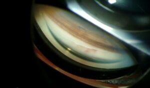
The surgeon dilates the pupil, uses a contact lens, and chooses a spot that covers the lesion with a margin.
Settings start low and rise in steps until the tissue shows only a faint gray change.
Then the surgeon treats the whole lesion in a confluent pattern for the planned time.
Repeat sessions at one to three months are common, so patients need clear follow-up plans (see choroidal melanoma).
Fundus Explorer Pro
Photograph the retinal findings described here with the phone already in your pocket — 22 D optics and built-in illumination in one handheld unit.
From Choroida — the team behind this siteTranspupillary Thermotherapy for Choroidal Melanoma
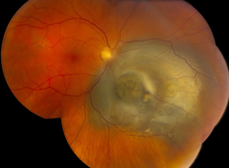
Early reports showed tumor control in small melanomas, mainly those thinner than 3 mm.
A large series of 256 consecutive cases reported good control but also recurrences, especially in tumors near the disc or with orange pigment.
Therefore, most centers now use it for small tumors away from the macula or as an adjunct after plaque therapy (see plaque brachytherapy for choroidal melanoma).
Transpupillary Thermotherapy in Retinoblastoma
Small peripheral retinoblastomas respond to heat, especially when combined with chemotherapy.
The heat increases the effect of carboplatin on tumor cells, so teams call the combination chemothermotherapy.
Larger or vitreous-seeding tumors need other treatments (see retinoblastoma).
Transpupillary Thermotherapy for Neovascular AMD
Studies in occult choroidal neovascularization found no clear gain in vision.
Thermal damage to the retina and the choroid often offset any benefit.
Anti-VEGF drugs replaced the method, so it has no current role in routine AMD care.
Complications
- Iris atrophy or pupil distortion after anterior treatment
- Retinal traction and macular folds after heavy treatment
- Vascular occlusion and focal field defects
- Cataract after treatment near the lens
Overheating is the main mechanism, and an experienced operator lowers the risk.
An Example Case
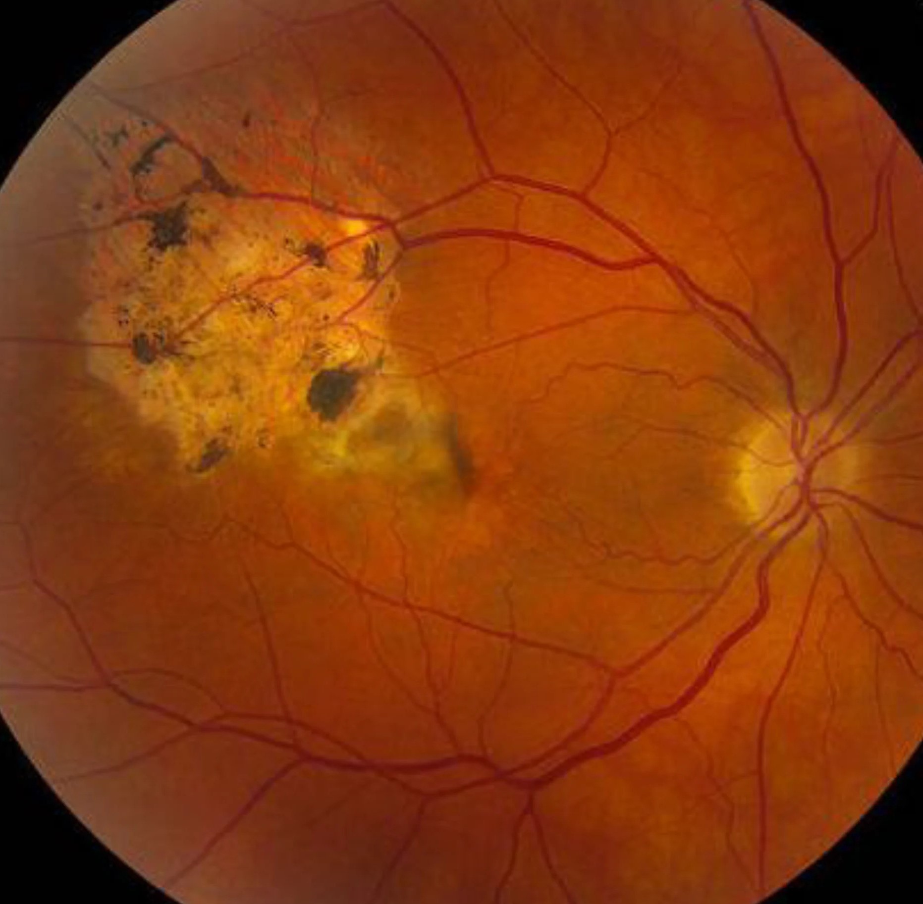
A 55-year-old woman has a 2.5 mm thick choroidal melanoma in the inferior periphery, 5 mm from the disc.
She prefers a plan that preserves central vision and accepts retreatment.
After staging, she receives transpupillary thermotherapy in two sessions.
The tumor flattens over six months, and she keeps 20/20 vision, with regular imaging.
Key Points
- Use it for small, thin tumors away from the macula and disc
- Avoid it for AMD, where anti-VEGF therapy is better
- Plan retreatment and long follow-up
- Refer pigmented or thick lesions for plaque or proton therapy
Questions Patients Ask
Patients ask whether the laser hurts, and most feel mild warmth or no pain under local anesthesia.
They also ask whether one session is enough, and the honest answer is that many need two or three.
Because the tumor may regrow, they need follow-up for years.
Pitfalls to Avoid
- Overheating the tissue, which causes retinal traction and vascular occlusion
- Treating thick tumors, where the heat does not reach the base
- Ignoring orange pigment or fluid, which signal growth risk
- Stopping follow-up after the first regression
Looking Ahead
Plaque and proton therapy now handle most melanomas that need treatment, so thermotherapy has become an adjunct.
Newer approaches combine heat with other agents, but the evidence is still limited.
However, the technique stays useful for small peripheral lesions in expert hands.
Practical Points
Take baseline photographs and ultrasound before treatment.
Then compare them at each visit, so that you can detect growth early.
Also coordinate with the ocular oncology team when features suggest growth.
Because metastatic risk depends on tumor biology, systemic surveillance continues even after local control.
Follow-Up After Treatment
After treatment, examine the eye every few weeks at first, then every few months.
Look for pigment changes, flattening, and fluid, since these signal response.
Because recurrence can appear years later, continue surveillance for life.
Also document visual field changes, which may reveal subtle scotomas from the treated area.


Document what you see
Two smartphone imaging tools built for everyday clinic use — one for the slit lamp, one for the fundus.
From Choroida — the team behind this siteReferences
- Oosterhuis JA, Journee-de Korver HG, Kakebeeke-Kemme HM, Bleeker JC. Transpupillary thermotherapy in choroidal melanomas. Arch Ophthalmol. 1995;113:315-321.
- Shields CL, Shields JA, Perez N, Singh AD, Cater J. Primary transpupillary thermotherapy for small choroidal melanoma in 256 consecutive cases: outcomes and limitations. Ophthalmology. 2002;109:225-234.
- Salmon JF. Kanski’s Clinical Ophthalmology: A Systematic Approach. 9th ed. Elsevier; 2020.
- American Academy of Ophthalmology. Basic and Clinical Science Course, Section 12: Retina and Vitreous. AAO; current edition.

Writes and reviews the clinical articles on Educate. Every post is checked against peer-reviewed sources before it goes live.