Pachychoroid spectrum disease describes a group of macular disorders linked by a thick, structurally abnormal choroid.
Members include central serous chorioretinopathy (CSCR), pachychoroid neovasculopathy and polypoidal choroidal vasculopathy.
Because they share imaging features, one map clarifies how they relate.
What the Pachychoroid Spectrum Describes
Freund and colleagues popularized the concept after describing pachychoroid pigment epitheliopathy in 2013.
The term groups diseases by choroidal phenotype.
However, it does not prove one identical cause.
Shared Imaging Features of the Pachychoroid Spectrum
Enhanced depth imaging or swept-source OCT shows the choroid best.
Several features recur across the group:
- Dilated Haller layer veins (pachyvessels)
- Thinning of Sattler layer and choriocapillaris over those veins
- Focal increase in choroidal thickness
- Choroidal hyperpermeability on indocyanine green angiography
- Overlying retinal pigment epithelium (RPE) changes
Fundus Explorer Pro
Photograph the retinal findings described here with the phone already in your pocket — 22 D optics and built-in illumination in one handheld unit.
From Choroida — the team behind this siteA Simple Pachychoroid Spectrum Map
First, the choroid develops pachyvessels without visible retinal damage.
Next, RPE changes may appear, and then subretinal fluid may follow.
Finally, chronic disease can produce type 1 neovascularization, sometimes with polyps.
However, patients need not pass through each stage in order.
Uncomplicated Pachychoroid
Some eyes show a thick choroid and pachyvessels without RPE disease or fluid.
Patients usually have normal vision, so the finding often emerges incidentally.
Its long-term risk is uncertain, so avoid labeling it active disease.
Pachychoroid Pigment Epitheliopathy
The examiner may see subtle RPE mottling, while the patient reports few symptoms.
OCT often shows small pigment epithelial detachments over pachyvessels.
Therefore, some authors regard this entity as a forme fruste of CSCR.
Central Serous Chorioretinopathy
CSCR adds serous macular detachment to the choroidal phenotype.
Patients describe blur, metamorphopsia or a central dim spot.
Corticosteroid exposure is a recognized risk factor, so ask about every route.
In chronic cases, autofluorescence may show descending tracks.
Pachychoroid Neovasculopathy
Pang and Freund described type 1 neovascularization arising over pachyvessels.
These eyes typically lack the soft drusen burden of neovascular AMD.
OCT often shows a shallow, irregular pigment epithelial detachment (double-layer sign).
Then, OCT angiography can reveal the flow network beneath the RPE.
This test helps because dye leakage may mimic chronic CSCR.
Polypoidal Choroidal Vasculopathy
Polypoidal lesions represent an aneurysmal form of type 1 neovascularization in many eyes.
Examination may show orange subretinal nodules, and hemorrhage can be large.
Indocyanine green angiography is the reference test for polyps.
However, not every polypoidal eye has a thick choroid.
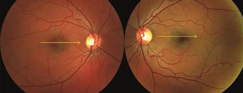
Peripapillary Pachychoroid Syndrome
This variant centers choroidal thickening around the optic disc.
OCT shows intraretinal and subretinal fluid near the disc, sometimes reaching the macula.
Mild disc edema can also create confusion with neuro-ophthalmic disease (see [peripapillary pachychoroid syndrome](peripapillary-pachychoroid-syndrome-pps-%d9%90a-full-study)).
Thickness Limits in Pachychoroid Spectrum
No universal thickness cutoff defines the diagnosis.
Many studies use values near 300 microns, yet healthy thickness varies widely.
Age, refractive error and time of day all influence the measurement.
Consequently, morphology carries more weight than one subfoveal number.
Workup for Pachychoroid Spectrum Eyes
Begin with dilated examination, then obtain structural OCT with deep choroidal imaging.
Next, add autofluorescence to document RPE damage and past fluid paths.
Also, OCT angiography helps when chronic fluid raises concern for hidden neovascularization.
Indocyanine green angiography then refines the diagnosis before photodynamic therapy.
Mimics of the Pachychoroid Spectrum
A thick choroid or subretinal fluid also occurs in other diseases.
Consider these first:
- Vogt-Koyanagi-Harada disease and posterior scleritis
- Uveal effusion syndrome
- Choroidal metastases or lymphoma
- Medication-related retinopathy, for example with MEK inhibitors
Treatment Across the Pachychoroid Spectrum
Treatment follows the active lesion, so the umbrella label does not dictate therapy.
For chronic CSCR, the PLACE trial favored half-dose photodynamic therapy over high-density subthreshold micropulse laser.
For polypoidal disease, EVEREST II found ranibizumab plus verteporfin therapy superior to ranibizumab monotherapy.
Anti-VEGF therapy targets neovascular activity, while it has limited value for fluid without neovascularization.
Photodynamic therapy can cause RPE atrophy or choroidal hypoperfusion, so discuss those risks first.
No treatment has been shown to reverse the choroidal phenotype.
Counseling in the Pachychoroid Spectrum
Explain that the term describes a choroidal pattern, and that one patient may never progress.
Advise prompt review for new distortion, blur or a central scotoma.
Fellow eyes also deserve imaging, because bilateral changes are common.
At follow-up, compare OCT fluid, RPE changes and any new sub-RPE flow.
Pitfalls to Avoid
- Do not diagnose disease from thickness alone
- Never assume chronic fluid lacks neovascularization without OCT angiography
- Do not overlook steroid creams, inhalers or injections


Document what you see
Two smartphone imaging tools built for everyday clinic use — one for the slit lamp, one for the fundus.
From Choroida — the team behind this siteReferences
- Warrow DJ, Hoang QV, Freund KB. Pachychoroid pigment epitheliopathy. Retina. 2013;33:1659-1672.
- Pang CE, Freund KB. Pachychoroid neovasculopathy. Retina. 2015;35:1-9.
- Cheung CMG, Lee WK, Koizumi H, et al. Pachychoroid disease. Eye. 2019;33:14-33.
- Phasukkijwatana N, Freund KB, Dolz-Marco R, et al. Peripapillary pachychoroid syndrome. Retina. 2018;38:1652-1667.
- van Dijk EHC, Fauser S, Breukink MB, et al. Half-dose photodynamic therapy versus high-density subthreshold micropulse laser treatment in patients with chronic central serous chorioretinopathy: the PLACE trial. Ophthalmology. 2018;125:1547-1555.
- Koh A, Lai TYY, Takahashi K, et al. Efficacy and safety of ranibizumab with or without verteporfin photodynamic therapy for polypoidal choroidal vasculopathy: a randomized clinical trial. JAMA Ophthalmol. 2017;135:1199-1206.

Writes and reviews the clinical articles on Educate. Every post is checked against peer-reviewed sources before it goes live.