Eyelid laceration is a common injury after falls, assaults, dog bites and blunt trauma.
Many of these cuts look simple at first glance.
However, a small skin wound can hide a margin defect, a torn canaliculus, a cut levator or an open globe.
Therefore, triage starts with the eye, then moves to the lid.
Examine the Globe First
Treat every lid wound as a possible globe injury until you exclude one.
First, check visual acuity, pupils and the anterior segment before you manipulate the lid.
A shallow chamber, peaked pupil, low pressure or uveal prolapse suggests rupture (see ruptured globe diagnosis).
In that case, avoid any pressure on the eye and place a rigid shield.
The open globe then takes priority, and lid repair waits.
History in Eyelid Laceration
The mechanism predicts the hidden damage.
For example, dog bites and hook injuries often tear the medial canthus and canaliculus.
Sharp objects can penetrate the septum and reach the orbit or globe.
Also ask about tetanus status, anticoagulants and the time since injury.
All-fit Slit-Lamp Adapter
Record and share exactly what you see at the slit lamp. One adapter fits any slit lamp or surgical microscope — and any smartphone.
From Choroida — the team behind this siteImaging Before Repair
Order a thin-cut orbital CT when you suspect a foreign body, fracture or deep penetration.
Avoid MRI until you exclude a metallic fragment.
However, imaging does not replace careful wound exploration.
Margin Involvement
Check whether the cut crosses the gray line and lash line.
A margin laceration needs precise alignment, because a small step leaves notching and lid malposition.
Simple skin closure across the margin often heals with a visible notch.
As a result, these wounds belong with an experienced surgeon.
Canalicular Injury in Eyelid Laceration
Any wound medial to the punctum can involve the canaliculus.
The lower canaliculus is involved more often than the upper one.
Dilate the punctum, probe gently and look for the probe in the wound.
Then plan repair over a stent (see canalicular repair technique).
Orbital Fat and the Septum
Yellow fat in the wound means the orbital septum is open.
Therefore, the injury is deeper than it looks.
In the upper lid, exposed fat also raises concern for levator damage.
Never suture the septum, because this can cause lid retraction and lagophthalmos.
Levator Damage in Eyelid Laceration
Ask the patient to look up, then measure levator excursion.
Ptosis after an upper lid cut may reflect swelling, hematoma or a torn aponeurosis.
Reattach the cut aponeurosis to the tarsus at primary repair when you find it.
Medial and Lateral Canthal Tendons
Pull the lid medially and laterally to test tendon integrity.
A rounded canthal angle or abnormal laxity suggests avulsion.
Repair should restore the posterior vector so the lid stays against the globe.
Tissue Loss
Eyelid tissue that looks lost is often only displaced or rolled.
Therefore, unfold and inspect every flap before you judge a defect.
Because lid tissue has an excellent blood supply, debride sparingly.
True full-thickness loss may need flaps or grafts (see margin reconstruction options).
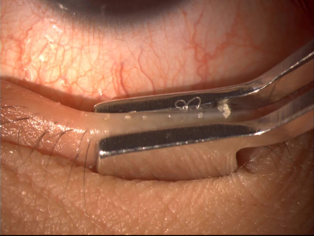
Which Eyelid Laceration Needs a Specialist
Superficial horizontal skin cuts away from the margin and canthus can often close in the emergency department.
However, refer these injuries to an oculoplastic or ophthalmic surgeon:
- Lacerations involving the lid margin
- Wounds medial to the punctum
- Exposed orbital fat or septal violation
- Ptosis or reduced levator function
- Canthal tendon avulsion
- Tissue loss or complex bites
- Any suspected globe injury or foreign body
Timing of Eyelid Laceration Repair
Repair the wound promptly once the patient is stable.
Many surgeons accept a short delay when the wound stays clean and moist.
However, edema and early fibrosis make canalicular ends harder to find over time.
Margin Repair Principles
First, align the gray line, lash line and tarsal edges.
Next, close the tarsus with partial-thickness absorbable sutures.
Then, place margin sutures with slight eversion to prevent notching.
Finally, close the skin and keep suture tails away from the cornea.
Anesthesia and Setting
Cooperative adults with simple wounds usually tolerate local anesthesia.
Children and complex injuries often need general anesthesia.
Antibiotics and Tetanus
Update tetanus prophylaxis according to local guidelines.
Irrigate bites and contaminated wounds thoroughly, and consider systemic antibiotics.
In addition, assess rabies risk after animal bites.
Counseling After Eyelid Laceration
Explain that swelling and bruising peak early and then settle.
Also warn about possible scarring, notching, ptosis or tearing.
Follow-Up and Late Problems
Check the cornea for exposure and suture rub at each visit.
Then assess lid closure, contour and tear drainage.
Late epiphora may signal canalicular obstruction.
Pitfalls to Avoid
- Do not repair the lid before you exclude an open globe
- Never close the orbital septum
- Do not miss a canalicular tear in medial wounds
- Avoid aggressive debridement of viable tissue
- Do not dismiss ptosis after an upper lid cut


Document what you see
Two smartphone imaging tools built for everyday clinic use — one for the slit lamp, one for the fundus.
From Choroida — the team behind this siteReferences
- American Academy of Ophthalmology. Basic and Clinical Science Course, Section 7: Oculofacial Plastic and Orbital Surgery. San Francisco: AAO; 2023.
- Bowling B. Kanski’s Clinical Ophthalmology: A Systematic Approach. 8th ed. Elsevier; 2016.
- Murchison AP, Bilyk JR. Canalicular laceration repair: an analysis of variables affecting success. Ophthal Plast Reconstr Surg. 2014;30:410-414.
- Wulc AE, Arterberry JF. The pathogenesis of canalicular laceration. Ophthalmology. 1991;98:1243-1249.
- Kuhn F, Morris R, Witherspoon CD, et al. A standardized classification of ocular trauma. Ophthalmology. 1996;103:240-243.
Test yourself
A few questions straight from this article.
-
Which assessment comes first when a patient presents with an eyelid laceration?
Every lid wound is treated as a possible globe injury until one is excluded, so acuity, pupils and the anterior segment are checked before the lid is manipulated. -
Which finding in a patient with an eyelid laceration suggests a ruptured globe?
A shallow chamber, peaked pupil, low pressure or uveal prolapse suggests rupture; the eye then gets a rigid shield with no pressure, and lid repair waits. -
Which imaging test should be ordered when an eyelid laceration raises suspicion of a foreign body or deep penetration?
Thin-cut orbital CT is ordered for a suspected foreign body, fracture or deep penetration, and MRI is avoided until a metallic fragment is excluded. -
What does yellow fat seen in an eyelid laceration indicate?
Yellow fat in the wound means the septum is open, so the injury is deeper than it looks; in the upper lid it also raises concern for levator damage. -
What can result from suturing the orbital septum during eyelid laceration repair?
The septum is never sutured because closing it can cause lid retraction and lagophthalmos. -
Which eyelid wound location should prompt a check for canalicular involvement?
Any wound medial to the punctum can involve the canaliculus, so the punctum is dilated and gently probed while looking for the probe in the wound. -
Why are margin sutures placed with slight eversion when repairing an eyelid margin laceration?
After the gray line, lash line and tarsal edges are aligned and the tarsus is closed, margin sutures go in with slight eversion to prevent notching. -
Why should debridement be sparing in eyelid laceration with apparent tissue loss?
Lid tissue that looks lost is often only displaced or rolled, and because it has an excellent blood supply, flaps are unfolded and inspected and debridement is kept sparing. -
What may late epiphora signal after repair of an eyelid laceration?
Follow-up checks lid closure, contour and tear drainage, and late epiphora may signal canalicular obstruction. -
Which eyelid laceration can often be closed in the emergency department without specialist referral?
Superficial horizontal skin cuts away from the margin and canthus can often close in the emergency department, whereas margin, medial, septal, levator and canthal injuries go to an oculoplastic or ophthalmic surgeon.

Writes and reviews the clinical articles on Educate. Every post is checked against peer-reviewed sources before it goes live.