Vitrectomy recovery usually takes weeks to months, and the pace depends on the disease and the tamponade.
A macular pucker with fluid fill often settles quickly, while a detachment repaired with long-acting gas takes far longer.
Therefore, this timeline is a general guide, and the surgeon’s instructions take priority.
What Sets the Vitrectomy Recovery Pace
Diagnosis, tamponade, lens status and macular condition shape the course.
For example, a chronic macula-off detachment may regain vision slowly despite perfect anatomical repair.
Diabetic eyes also carry a risk of recurrent vitreous hemorrhage.
Day One: The First Vitrectomy Recovery Visit
The examiner checks vision, intraocular pressure, wounds, anterior chamber reaction and retinal attachment.
Pressure may run high because of inflammation, retained viscoelastic or an overfilled bubble.
However, sutureless small-gauge wounds can also leak and cause transient hypotony.
Fundus Explorer Pro
Photograph the retinal findings described here with the phone already in your pocket — 22 D optics and built-in illumination in one handheld unit.
From Choroida — the team behind this siteWeek One: Normal Symptoms
Redness, lid swelling, tearing and a scratchy sensation are common in this period.
Patients often report mild ache, which usually responds to simple analgesia.
Vision may look very blurred, especially behind a gas bubble or with dilating drops on board.
In addition, subconjunctival hemorrhage can look alarming, although it clears over about two weeks without treatment.
Drops During Vitrectomy Recovery
Most regimens combine a topical antibiotic, a corticosteroid and sometimes a cycloplegic.
The steroid controls inflammation, while the cycloplegic relieves ciliary spasm and discomfort.
However, steroids can raise pressure in responders, so repeat tonometry during the taper.
Face-Down and Head Positioning
Gas floats, so positioning places the bubble against the target break or macular hole.
Schedules differ by break location and surgeon, so patients need a written position and duration.
Also, patients with gas should avoid sleeping flat on their back, because the bubble can press on the lens and iris.
The Gas Bubble Timeline
Absorption time depends on the gas and its concentration (see tamponade choice).
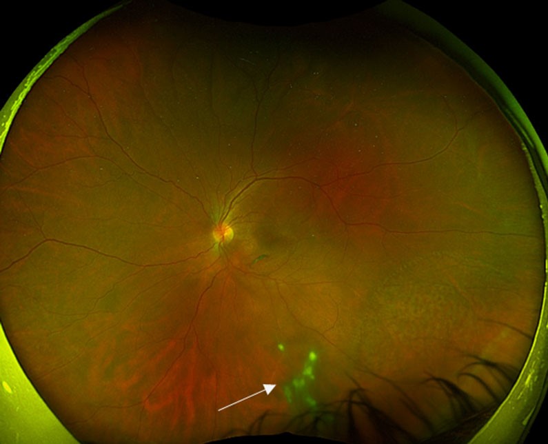
Air generally clears within about a week, SF6 within a few weeks, and C3F8 over roughly two months.
Patients first see a wobbling dark line, which shrinks into a small circle in lower vision.
Finally, the bubble often breaks into tiny beads before disappearing.
Gas Safety During Vitrectomy Recovery
Gas expands at lower ambient pressure and with nitrous oxide anesthesia.
Simulated flight testing showed pressure rises in gas-filled eyes, and nitrous oxide exposure has caused permanent vision loss.
Therefore, patients must avoid flying and high-altitude travel until the surgeon confirms the gas has cleared.
They should also warn any anesthetist, and many surgeons supply a warning wristband.
Silicone Oil Follows a Different Path
Silicone oil does not expand at altitude, so flying is generally permitted.
However, vision often stays limited, and most eyes need a second operation for removal.
Meanwhile, follow-up tracks pressure, emulsification and corneal changes.
Weeks Two to Four: Activity in Vitrectomy Recovery
Reading and watching television do not harm the eye, and light walking is usually fine.
Surgeons commonly restrict heavy lifting, strenuous exercise and swimming for a few weeks, although evidence for exact limits is sparse.
Desk workers often return earlier than manual workers.
Driving depends on binocular vision, legal standards and the presence of a bubble.
Weeks Four to Eight: Vision Returns
As gas clears, the clinician can image the macula with OCT and confirm hole closure.
Persistent blur may reflect macular edema, an epiretinal membrane or photoreceptor damage.
Next, defer new glasses until the refraction stabilizes.
Months Three to Twelve of Vitrectomy Recovery
Distortion and acuity can keep improving slowly for many months after macular surgery.
However, nuclear sclerosis commonly progresses in phakic eyes, particularly in older patients (see cataract after vitrectomy).
Therefore, vision that improves and then gradually fades often points to the lens.
Warning Signs After Vitrectomy
Patients should call the same day for any of these:
- Worsening pain or increasing redness
- Falling vision or a new dark curtain
- New flashes or a shower of floaters
- Purulent discharge
- Severe eye pain with nausea or vomiting
Urgent Causes to Exclude
Endophthalmitis is uncommon after small-gauge and 20-gauge surgery, yet it threatens vision quickly.
Also, pressure spikes can injure the optic nerve, so check tonometry at any urgent visit.
Finally, new field loss suggests redetachment, which often involves proliferative vitreoretinopathy (see recurrent detachment).
Counseling for Vitrectomy Recovery
Explain early that visual outcome depends largely on the preoperative macula.
Successful anatomy does not guarantee sharp vision, so set realistic goals before surgery.
Pitfalls to Avoid
- Do not clear a patient to fly without confirming gas absorption
- Avoid nitrous oxide in any eye with residual gas
- Do not blame cataract before examining the macula with OCT


Document what you see
Two smartphone imaging tools built for everyday clinic use — one for the slit lamp, one for the fundus.
From Choroida — the team behind this siteReferences
- Mills MD, Devenyi RG, Lam WC, et al. An assessment of intraocular pressure rise in patients with gas-filled eyes during simulated air flight. Ophthalmology. 2001;108:40-44.
- Seaberg RR, Freeman WR, Goldbaum MH, Manecke GR. Permanent postoperative vision loss associated with expansive gas in the setting of nitrous oxide anesthesia. Anesthesiology. 2002;97:1309-1310.
- Scott IU, Flynn HW Jr, Dev S, et al. Endophthalmitis after 25-gauge and 20-gauge pars plana vitrectomy: incidence and outcomes. Retina. 2008;28:138-142.
- American Academy of Ophthalmology. Basic and Clinical Science Course, Section 12: Retina and Vitreous. San Francisco: AAO.
- Schachat AP, Sadda SR, Hinton DR, et al. Ryan’s Retina. 6th ed. Philadelphia: Elsevier; 2018.

Writes and reviews the clinical articles on Educate. Every post is checked against peer-reviewed sources before it goes live.