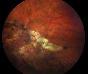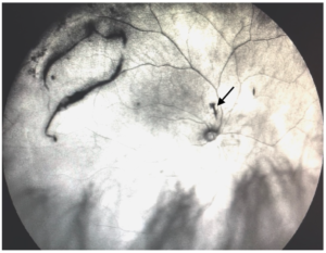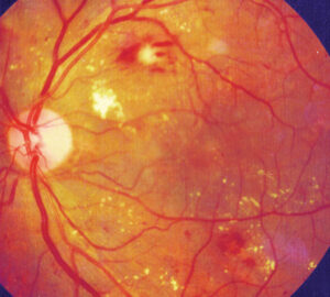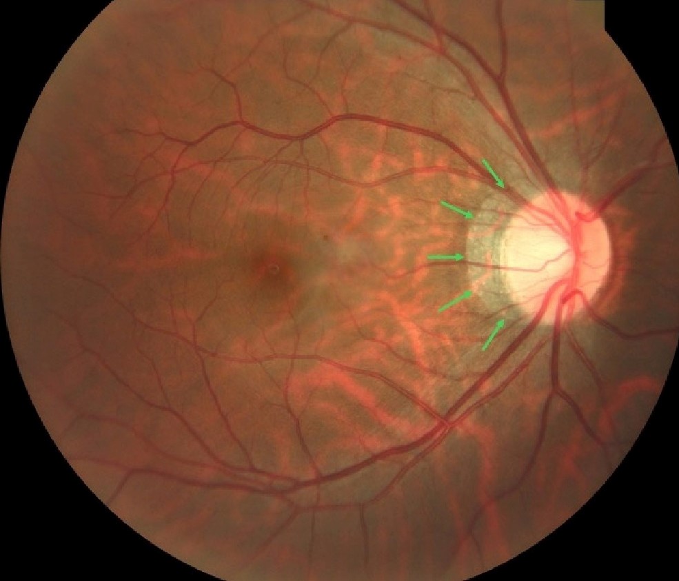
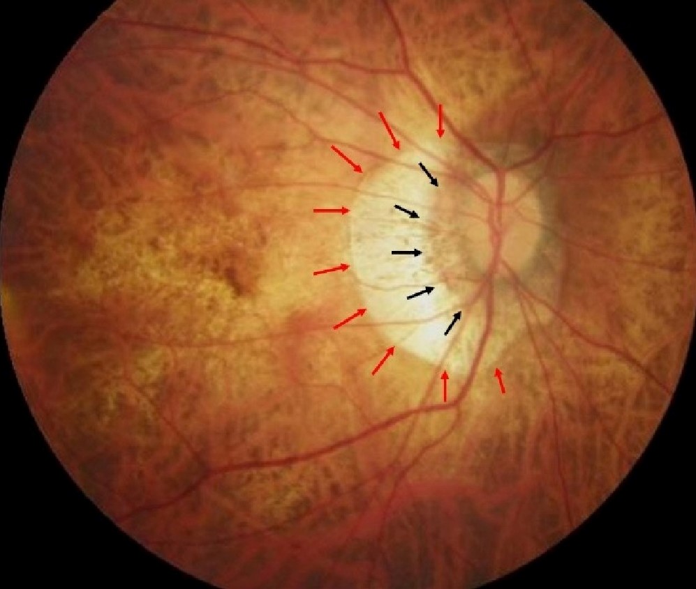
Peripapillary atrophy is a crescent or ring of chorioretinal thinning around the optic disc.
It occurs in normal eyes, but it enlarges in myopia, glaucoma, and some other diseases.
Understanding its zones helps to interpret the optic disc and the risk of progression.
Zones of Peripapillary Atrophy
Jonas and colleagues described two zones in the 1980s.
- Alpha zone: the outer area with irregular pigment and thinning of the retinal pigment epithelium
- Beta zone: the inner area with complete loss of RPE and visible sclera and large choroidal vessels
Later work defined a gamma zone, which is the region of peripapillary tissue without Bruch membrane, seen mainly in myopic eyes.
Kubota and colleagues correlated these zones with histology (see pathologic myopia classification).
Peripapillary Atrophy in Glaucoma
The beta zone is larger and more frequent in glaucoma than in healthy eyes.
It often lies next to areas of neuroretinal rim loss and retinal nerve fiber layer defects.
Jonas reviewed these data and noted that the extent of beta zone correlates with the degree of glaucomatous damage in many patients.
However, myopia also enlarges the beta zone, so interpret the sign with caution.
Fundus Explorer Pro
Photograph the retinal findings described here with the phone already in your pocket — 22 D optics and built-in illumination in one handheld unit.
From Choroida — the team behind this sitePeripapillary Atrophy in Myopia
In myopia, peripapillary atrophy enlarges as the eye elongates, and the temporal crescent is common.
Dai and colleagues reported that gamma zone widens with axial length, while the beta zone relates more closely to glaucoma.
This distinction matters, because a large crescent in a highly myopic eye does not always mean glaucoma (see tilted disc syndrome).
Other Associations
- Age-related macular degeneration, with larger zones in some studies
- Posterior staphyloma and pathologic myopia
- Optic disc anomalies such as tilted discs
- Normal eyes, where a small alpha zone is common
Imaging of Peripapillary Atrophy
Color photographs show the extent of the crescent.
OCT shows the thinning of the RPE and Bruch membrane, and it helps to separate beta and gamma zones.
Fundus autofluorescence shows loss of signal in the beta zone, and OCT angiography may show reduced capillary density.
Clinical Use
Record the size and the location of the crescent at baseline.
Compare serial photographs for enlargement, which may signal progression of glaucoma or myopia.
Use the sign together with pressure, fields, and OCT of the nerve fiber layer.
An Example Case
A 62-year-old man has ocular hypertension and a large temporal crescent around the right disc.
OCT of the retinal nerve fiber layer shows a localized defect next to the crescent.
Photographs show that the beta zone has enlarged over two years, and the cup has deepened in the same sector.
The clinician diagnoses early glaucoma, and starts treatment.
Questions Patients Ask
Patients ask whether the crescent is dangerous, and in many people it is a normal feature.
They also ask whether it will grow, and in myopia it may grow with the length of the eye.
Because the sign is not specific, explain that the doctor uses it together with other tests.
Pitfalls to Avoid
- Calling a myopic crescent glaucoma without other evidence
- Overlooking progression because photographs were not saved
- Mixing alpha and beta zones in the chart
- Forgetting to check the fellow eye
Looking Ahead
Automated OCT measures of zones may standardize assessment.
Also, studies may clarify how gamma and beta zones relate to glaucoma risk in myopia.
Therefore, interpretation may become more precise.
Practical Points
Photograph the disc at baseline, and repeat every year or two.
Then compare the crescent size and the rim in each sector.
Also use OCT to confirm structural changes.
Peripapillary Atrophy and Axial Length
Axial length affects the size of the zones, so record it when available.
Because high myopia enlarges the crescent, adjust expectations.
Then use the location of the crescent relative to rim loss as a guide.
Distinguishing Glaucoma From Myopia
Look at the rim, the nerve fiber layer, and the visual field together.
Because myopia enlarges the crescent without glaucoma, use sector-specific rim loss and field defects as clues.
Also compare with prior photographs.
Then use OCT of the ganglion cell layer to support the diagnosis.
Patient Information
Explain that a crescent is common and often benign.
Also tell patients that regular follow-up protects vision when glaucoma risk exists.
Documentation Tips
Use the same camera and field for serial photographs.
Because small changes matter, mark the disc margin and the crescent edge.
Also record the axial length when it is available.
For the latest studies, see the PubMed results for peripapillary atrophy.


Document what you see
Two smartphone imaging tools built for everyday clinic use — one for the slit lamp, one for the fundus.
From Choroida — the team behind this siteReferences
- Jonas JB, Nguyen XN, Gusek GC, Naumann GO. Parapapillary chorioretinal atrophy in normal and glaucoma eyes. I. Morphometric data. Invest Ophthalmol Vis Sci. 1989;30:908-918.
- Jonas JB. Clinical implications of peripapillary atrophy in glaucoma. Curr Opin Ophthalmol. 2005;16:84-88.
- Dai Y, Jonas JB, Huang H, et al. Microstructure of parapapillary atrophy: beta zone and gamma zone. Invest Ophthalmol Vis Sci. 2013;54:2013-2018.
- American Academy of Ophthalmology. Basic and Clinical Science Course, Section 10: Glaucoma. AAO; current edition.
Test yourself
A few questions straight from this article.
-
Which description best defines peripapillary atrophy?
Peripapillary atrophy is a crescent or ring of chorioretinal thinning around the optic disc; it occurs in normal eyes but enlarges in myopia and glaucoma. -
Which feature defines the alpha zone of peripapillary atrophy?
The alpha zone is the outer area, with irregular pigment and thinning of the retinal pigment epithelium; a small alpha zone is common in normal eyes. -
Which feature defines the beta zone of peripapillary atrophy?
The beta zone is the inner area, where the RPE is completely lost so the sclera and large choroidal vessels become visible. -
The gamma zone of peripapillary atrophy is defined by the absence of which structure?
The gamma zone is peripapillary tissue without Bruch membrane, seen mainly in myopic eyes. -
In glaucoma, the beta zone of peripapillary atrophy typically lies next to which findings?
The beta zone is larger and more frequent in glaucoma and often lies beside areas of rim loss and retinal nerve fiber layer defects. -
Which peripapillary atrophy zone widens with axial length in myopic eyes?
Dai and colleagues found that the gamma zone widens with axial length, while the beta zone relates more closely to glaucoma. -
How does the beta zone of peripapillary atrophy appear on fundus autofluorescence?
Fundus autofluorescence shows loss of signal in the beta zone, where the RPE is absent. -
Which imaging test helps separate the beta zone from the gamma zone of peripapillary atrophy?
OCT shows the thinning of the RPE and Bruch membrane, which allows the beta and gamma zones to be told apart. -
In a myopic eye with a large peripapillary crescent, which findings best support coexisting glaucoma?
Myopia enlarges the crescent without glaucoma, so sector-specific rim loss and field defects, with nerve fiber layer OCT, are the clues to glaucoma. -
How often should optic disc photographs be repeated when monitoring peripapillary atrophy?
Photograph the disc at baseline and repeat every year or two, comparing crescent size and rim in each sector; enlargement may signal progression.

Writes and reviews the clinical articles on Educate. Every post is checked against peer-reviewed sources before it goes live.