Vitrectomy for diabetic retinopathy removes blood and fibrovascular tissue from the vitreous cavity and relieves traction on the retina.
Surgeons reserve it for the complications of proliferative disease that laser and injections alone cannot handle.
The timing of surgery matters, and the availability of anti-VEGF drugs has changed how the procedure is planned.
When Is Vitrectomy for Diabetic Retinopathy Needed?
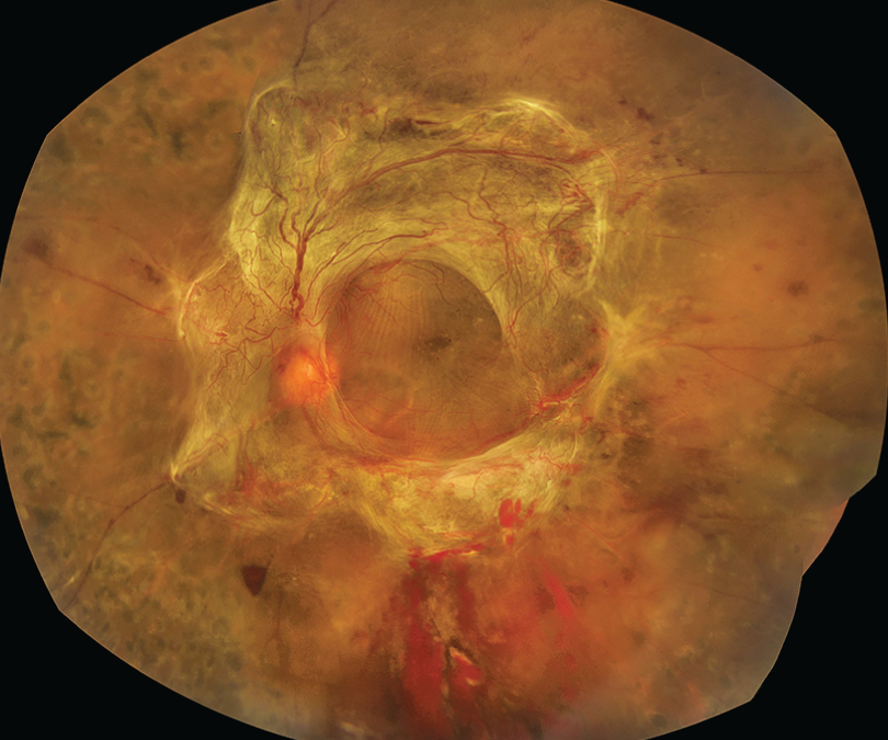
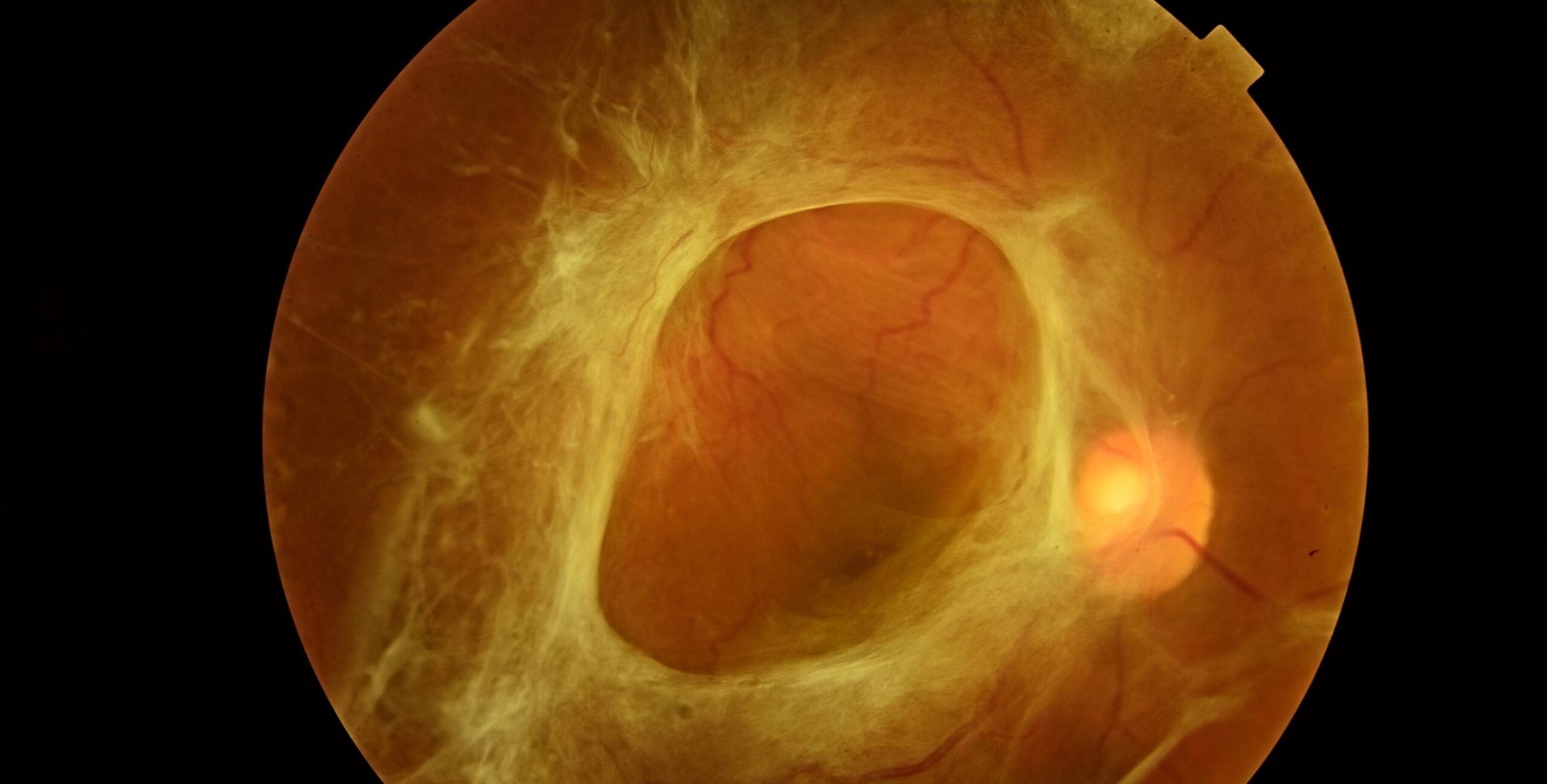
Proliferative retinopathy produces fibrovascular tissue that adheres to the posterior hyaloid and the retina.
As the vitreous contracts, this tissue pulls on the retina, tears vessels, and bleeds.
Surgery clears media opacities that prevent laser treatment and removes the scaffold of traction.
Most patients who need vitrectomy have already had laser, injections, or both.
The procedure is a step after those measures.
Indications of Vitrectomy for Diabetic Retinopathy
- Nonclearing vitreous hemorrhage: commonly after one to three months of observation, or sooner in type 1 diabetes or if the other eye is poor (see vitreous hemorrhage)
- Tractional retinal detachment involving or threatening the macula (see tractional retinal detachment)
- Combined tractional and rhegmatogenous detachment (see combined detachment)
- Dense premacular subhyaloid hemorrhage that does not clear
- Severe fibrovascular proliferation with progressive traction
- Diabetic macular edema with vitreomacular traction or a taut posterior hyaloid in selected cases
- Anterior segment neovascularization with hemorrhage that blocks laser
Fundus Explorer Pro
Photograph the retinal findings described here with the phone already in your pocket — 22 D optics and built-in illumination in one handheld unit.
From Choroida — the team behind this siteVitrectomy for Diabetic Retinopathy: Timing
The Diabetic Retinopathy Vitrectomy Study found that early vitrectomy for severe vitreous hemorrhage gave better visual outcomes in type 1 diabetes, with less benefit in type 2.
Today, many surgeons observe a fresh hemorrhage for a few weeks while giving anti-VEGF drugs, and operate if it does not clear.
Operate promptly on a tractional detachment threatening the macula, because delay allows the detachment to progress.
Preoperative Anti-VEGF
Injection of an anti-VEGF drug a few days before surgery can regress new vessels, reduce bleeding at surgery, and make membrane dissection easier.
The interval is usually 3 to 7 days, because a longer interval can cause rapid contraction of the fibrovascular tissue and worsening traction, a recognized event sometimes called the crunch phenomenon (see crunch syndrome).
Patients with known tractional detachment should be treated with caution, and the surgery should not be delayed after injection.
Surgical Steps
The surgeon performs a core vitrectomy, then separates the posterior hyaloid and removes the fibrovascular membranes by peeling, segmentation, and delamination.
The surgeon applies endolaser panretinal photocoagulation to the retina to reduce the ischemic drive.
Also, the surgeon controls bleeding by raising the infusion pressure, using diathermy, or injecting anti-VEGF.
Clinicians use tamponade with gas or silicone oil when there are retinal breaks or a detachment (see silicone oil tamponade and vitreous substitutes).
Outcomes and Complications of Vitrectomy for Diabetic Retinopathy
Visual outcomes depend on the preoperative status of the macula, the degree of ischemia, and the presence of detachment.
Many patients improve substantially after clearance of a hemorrhage, but eyes with extensive macular ischemia or a long-standing macular detachment may not.
Complications include recurrent vitreous hemorrhage, iatrogenic retinal breaks, cataract, neovascular glaucoma, and proliferative vitreoretinopathy (see vitrectomy complications).
Counsel patients that surgery treats the complication and not the underlying disease, so laser, injections, and systemic control continue after the operation.
Planning the Operation
Before surgery, review the systemic status: glucose control, blood pressure, kidney function, and anticoagulant use.
Document the status of the fellow eye, because a patient with only one useful eye needs particular care in planning.
Ultrasound is needed when hemorrhage blocks the view, to look for a tractional or combined detachment and to plan the approach (see B-scan ultrasonography).
Small-gauge vitrectomy is widely used and offers faster recovery.
The choice of gauge depends on the surgeon and the case.
Anesthesia and Positioning
Surgeons do most operations under local anesthesia with sedation, and use general anesthesia when the patient cannot lie still or the case is expected to be long.
After surgery with gas, the surgeon asks the patient to keep a particular head position.
Those with silicone oil may need positioning too, so explain the plan beforehand.
Postoperative Care
Review the patient the next day and again within a week, with attention to pressure, hemorrhage, and wound integrity.
Continue panretinal photocoagulation in the clinic if the periphery was not fully treated, and consider anti-VEGF injections if neovascularization persists.
Recurrent hemorrhage is common in the first weeks, and it often clears without further surgery.
Counsel the patient on realistic visual expectations, and refer for low-vision services if vision remains limited.
For the latest studies, see the PubMed results for vitrectomy for diabetic retinopathy.


Document what you see
Two smartphone imaging tools built for everyday clinic use — one for the slit lamp, one for the fundus.
From Choroida — the team behind this siteReferences
- Diabetic Retinopathy Vitrectomy Study Research Group. Early vitrectomy for severe vitreous hemorrhage in diabetic retinopathy: two-year results of a randomized trial. DRVS report 2. Arch Ophthalmol. 1985;103:1644-1652.
- Diabetic Retinopathy Vitrectomy Study Research Group. Early vitrectomy for severe vitreous hemorrhage in diabetic retinopathy: four-year results of a randomized trial. DRVS report 5. Arch Ophthalmol. 1990;108:958-964.
- Ryan SJ, Schachat AP, Wilkinson CP, et al, eds. Retina. 6th ed. Elsevier; 2018.
- American Academy of Ophthalmology. Basic and Clinical Science Course, Section 12: Retina and Vitreous. AAO; current edition.

Writes and reviews the clinical articles on Educate. Every post is checked against peer-reviewed sources before it goes live.