
Thyroid eye disease lid signs are the eyelid findings of Graves orbitopathy, and they are often the earliest and most visible signs.
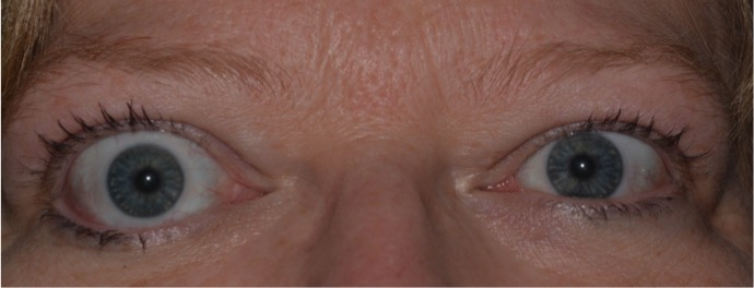
Upper eyelid retraction is the most common feature, and several named signs describe its variations.
Knowing the signs and what they mean helps to recognize the disease and to follow its activity.
Why the Eyelids Are Affected
The upper lid is elevated by the levator muscle and by Müller muscle, which is sympathetically innervated.
In Graves disease, the Müller muscle is overstimulated, and the levator and Müller muscles become inflamed and fibrotic.
In addition, tethering of the inferior rectus restricts downgaze, which causes the superior rectus and levator complex to overact in compensation.
Proptosis adds to the appearance by pushing the lids back.
Early retraction can improve when the thyroid state is controlled, while established fibrotic retraction needs surgery.
The Named Thyroid Eye Disease Lid Signs
- Dalrymple sign: upper eyelid retraction that exposes the sclera above the limbus
- von Graefe sign: lid lag, in which the upper lid fails to follow the globe smoothly on downgaze
- Kocher sign: a staring or frightened look caused by retraction on attempted fixation
- Stellwag sign: infrequent blinking
- Boston sign: jerky movements of the upper lid on downgaze
- Gifford sign: difficulty in everting the upper lid
- Rosenbach sign: tremor of the closed lids
- Joffroy sign: absence of forehead wrinkling on upgaze
- Möbius sign: poor convergence (see convergence insufficiency)
Several are of historical interest, and the first two are the most useful clinically.
All-fit Slit-Lamp Adapter
Record and share exactly what you see at the slit lamp. One adapter fits any slit lamp or surgical microscope — and any smartphone.
From Choroida — the team behind this siteHow to Examine
Position the patient with the eyes in primary gaze, and measure the margin reflex distance, the vertical palpebral fissure, and the amount of scleral show above and below the limbus.
Ask the patient to look slowly downward, and observe the lid movement for lag.
Look for lagophthalmos on gentle lid closure, check the cornea for exposure, and measure proptosis with an exophthalmometer.
Assess activity with the clinical activity score, which includes pain, redness, swelling, and change in function.
Examine the eye movements and the optic nerve function, since compressive optic neuropathy is the most urgent complication.
Differential Diagnosis
Other causes of eyelid retraction include dorsal midbrain lesions, aberrant regeneration, previous surgery, and contralateral ptosis (see eyelid retraction causes).
A patient with isolated retraction and no thyroid disease needs a broader evaluation.
Management
Keep the thyroid state stable, stop smoking, and use lubricants and ointments for corneal protection (see exposure keratopathy).
In active disease, treatments include selenium for mild disease, intravenous glucocorticoids, orbital radiotherapy, and teprotumumab, with the guidelines of the European Group on Graves’ Orbitopathy as a reference.
In the inactive phase, rehabilitative surgery is done in order: orbital decompression, strabismus surgery, and then eyelid surgery.
Eyelid procedures include recession of Müller muscle and the levator, spacer grafts for the lower lid, and blepharoplasty for fat and skin excess (see blepharoptosis surgical techniques).
Small amounts of triamcinolone or botulinum toxin can temporarily lower the lid.
Explain to the patient that the appearance can improve, and that timing and sequence matter.
Activity and Severity
The lid signs help to assess the severity, but the activity of the disease is judged by inflammatory signs: pain, redness, eyelid and conjunctival swelling, and recent change in function.
An active, inflamed orbit responds to anti-inflammatory therapy, while an inactive orbit with a stable lid position needs rehabilitation.
Document the lid measurements at each visit, so that change can be recognized, and use photographs to supplement the record.
Corneal Protection
Lid retraction and proptosis increase the area of exposed ocular surface, and the cornea may develop punctate keratopathy, ulcers, or infection.
Use preservative-free lubricants in the day and ointment at night, and consider moisture goggles.
Teach patients to recognize the warning signs of corneal trouble: pain, redness, and a decrease in vision.
Urgent surgery or lid taping may be needed for severe exposure.
Smoking and Thyroid Control
Smoking is a strong risk factor for the development and severity of the disease, and quitting improves outcomes.
Maintain euthyroidism with the help of the endocrinologist, because both hypothyroidism and hyperthyroidism worsen the eye disease.
Radioactive iodine can worsen the orbitopathy in some patients, and steroid prophylaxis is used in at-risk patients.
Patient Counselling
Many patients are concerned about their appearance, and the lid signs are conspicuous.
Explain that the early changes may recede, that treatment is available for each stage, and that psychological support is a legitimate part of care.
Encourage regular follow-up and early report of double vision, color changes, or visual loss, since these may signal optic neuropathy.


Document what you see
Two smartphone imaging tools built for everyday clinic use — one for the slit lamp, one for the fundus.
From Choroida — the team behind this siteReferences
- Bartley GB, Gorman CA. Diagnostic criteria for Graves’ ophthalmopathy. Am J Ophthalmol. 1995;119:792-795.
- Bartalena L, Kahaly GJ, Baldeschi L, et al. The 2021 European Group on Graves’ Orbitopathy (EUGOGO) clinical practice guidelines for the medical management of Graves’ orbitopathy. Eur J Endocrinol. 2021;185:G43-G67.
- Bartley GB. The differential diagnosis and classification of eyelid retraction. Ophthalmology. 1996;103:168-176.
- American Academy of Ophthalmology. Basic and Clinical Science Course, Section 7: Oculofacial Plastic and Orbital Surgery. AAO; current edition.

Writes and reviews the clinical articles on Educate. Every post is checked against peer-reviewed sources before it goes live.


