
Staphylococcal marginal keratitis produces peripheral corneal infiltrates in patients with chronic staphylococcal blepharitis, and the essential concept to understand is that this is a hypersensitivity reaction to staphylococcal exotoxins and cell wall components, not a direct bacterial invasion of the cornea, which is exactly why its management differs meaningfully from that of an infectious bacterial keratitis despite a superficially similar appearance.
The Immune Mechanism
Chronic staphylococcal colonization of the eyelid margins, common in blepharitis, exposes the peripheral cornea to ongoing low-level antigenic stimulation from staphylococcal exotoxins and cell wall components.
Over time, this produces a localized immune hypersensitivity response in the peripheral cornea, generating characteristic infiltrates that represent an immune reaction to bacterial products rather than a site of active bacterial invasion within the corneal tissue itself, a distinction that is central to understanding why these lesions typically remain sterile even though they arise directly from a bacterial colonization process.
Clinical Features
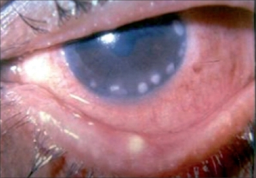
- One or more small, typically peripheral, corneal infiltrates, often multiple and sometimes bilateral
- A characteristic “clear interval” between the infiltrate and the limbus, a small zone of clear cornea separating the lesion from the peripheral vascular arcade, a helpful distinguishing clinical sign
- Mild to moderate pain, redness, and foreign body sensation, generally less severe than the pain typical of an infectious bacterial keratitis
- Signs of chronic blepharitis, including lid margin erythema, telangiectasia, and collarettes at the lash base, are almost always present and support the diagnosis
- The overlying epithelium may be intact or show only minimal, if any, staining, distinguishing it from an infectious ulcer where epithelial breakdown over the infiltrate is typical
All-fit Slit-Lamp Adapter
Record and share exactly what you see at the slit lamp. One adapter fits any slit lamp or surgical microscope — and any smartphone.
From Choroida — the team behind this siteDistinguishing From Infectious Bacterial Keratitis
This distinction has real management implications, since infectious keratitis needs aggressive antimicrobial treatment while marginal keratitis is managed primarily as an inflammatory, hypersensitivity-driven process.
- The characteristic clear interval between the lesion and the limbus favors marginal keratitis, while infectious infiltrates more often extend to or arise directly at the limbus
- Marginal keratitis lesions are typically smaller and less densely infiltrated, with less overlying epithelial defect, compared with a typical infectious infiltrate
- The presence of significant chronic blepharitis supports marginal keratitis, though blepharitis and infectious keratitis can coexist, so this clue is supportive rather than absolute
- Pain and anterior chamber reaction are generally milder in marginal keratitis than in an infectious process of comparable infiltrate size
Diagnosis
Diagnosis is largely clinical, based on the characteristic appearance, location, and clear interval, combined with evidence of chronic blepharitis.
Culture is not typically necessary for a classic presentation, though it is considered when the diagnosis is uncertain, the lesion is atypical in location or appearance, or when the response to treatment is not as expected, since missing a true infectious keratitis would have more serious consequences.
Management
Treating the Underlying Blepharitis
Since staphylococcal marginal keratitis arises from chronic lid margin colonization, addressing the underlying blepharitis, through lid hygiene and, when needed, topical or oral antibiotic treatment directed at the lid margin, is a central part of both treating the current episode and preventing recurrence (see blepharitis).
Topical Corticosteroids
Because the lesion is primarily an immune hypersensitivity reaction, low-potency topical corticosteroids are often used to reduce the inflammatory infiltrate and speed resolution, generally combined with or following a course of topical antibiotic treatment to address the underlying bacterial colonization driving the hypersensitivity response.
Combination Approach
A typical treatment approach combines a topical antibiotic-corticosteroid combination or sequential antibiotic followed by corticosteroid treatment, alongside ongoing lid hygiene measures, tailored to the severity of the presentation and the degree of underlying blepharitis.
Prognosis
Staphylococcal marginal keratitis generally responds well to appropriate treatment, with resolution of the infiltrates over one to a few weeks, though recurrence is common in patients with poorly controlled chronic blepharitis, underscoring the importance of ongoing lid hygiene as a preventive measure rather than treatment directed only at acute episodes as they occur.
Recognizing the hypersensitivity nature of this condition, distinct from direct infection, supports both more accurate diagnosis and a treatment approach, including judicious corticosteroid use, that would not be appropriate for a true infectious corneal ulcer.


Document what you see
Two smartphone imaging tools built for everyday clinic use — one for the slit lamp, one for the fundus.
From Choroida — the team behind this siteReferences
- Ficker L, Ramakrishnan M, Seal D, Wright P. Role of cell-mediated immunity to staphylococci in blepharitis. Am J Ophthalmol. 1991;111:473-479.
- Mondino BJ. Inflammatory diseases of the peripheral cornea. Ophthalmology. 1988;95:463-472.
- Smolin G, Okumoto M. Staphylococcal blepharitis. Arch Ophthalmol. 1977;95:812-816.
- Bowling B. Kanski’s Clinical Ophthalmology: A Systematic Approach. 8th ed. Edinburgh: Elsevier; 2016.
Test yourself
A few questions straight from this article.
-
What drives the peripheral corneal infiltrates of staphylococcal marginal keratitis?
Chronic lid margin colonisation exposes the peripheral cornea to staphylococcal exotoxins and cell wall components, producing a localised hypersensitivity response rather than bacterial invasion of the cornea. -
Which slit-lamp sign favours staphylococcal marginal keratitis over an infectious corneal infiltrate?
A small zone of clear cornea separates the infiltrate from the peripheral vascular arcade, whereas infectious infiltrates more often extend to or arise at the limbus. -
Which lid margin findings almost always accompany staphylococcal marginal keratitis?
Signs of chronic blepharitis, including lid margin erythema, telangiectasia and collarettes at the lash base, are almost always present and support the diagnosis. -
How does the epithelium overlying a staphylococcal marginal keratitis infiltrate typically appear?
The overlying epithelium may be intact or show minimal staining at most, unlike an infectious ulcer where epithelial breakdown over the infiltrate is typical. -
Compared with infectious bacterial keratitis of similar infiltrate size, how do pain and anterior chamber reaction behave?
Pain and anterior chamber reaction are generally milder in marginal keratitis than in an infectious process with an infiltrate of comparable size. -
When should corneal culture be taken in suspected staphylococcal marginal keratitis?
Culture is unnecessary for a classic presentation but is considered when the diagnosis is uncertain, the lesion is atypical, or treatment response is not as expected, since missing true infectious keratitis is more serious. -
Why is treating the underlying blepharitis central to managing staphylococcal marginal keratitis?
The condition arises from chronic lid margin colonisation, so lid hygiene and, when needed, antibiotic treatment directed at the lid margin both treat the episode and prevent recurrence. -
What is the purpose of low-potency topical corticosteroids in staphylococcal marginal keratitis?
Because the lesion is primarily an immune hypersensitivity reaction, low-potency topical corticosteroids reduce the inflammatory infiltrate and speed resolution, combined with or following topical antibiotic treatment. -
Over what period do the infiltrates of staphylococcal marginal keratitis typically resolve with appropriate treatment?
The condition generally responds well to appropriate treatment, with the infiltrates resolving over one to a few weeks. -
Which factor makes recurrent staphylococcal marginal keratitis most likely?
Recurrence is common where chronic blepharitis stays poorly controlled, which is why ongoing lid hygiene matters as prevention rather than treating only acute episodes.

Writes and reviews the clinical articles on Educate. Every post is checked against peer-reviewed sources before it goes live.


