
Blepharoptosis surgical repair corrects an abnormally low eyelid position through one of several distinct surgical approaches, and the choice among them depends heavily on how well the levator muscle itself is still functioning, discussed more broadly in relation to ptosis in its own dedicated article on this site.
Rather than a single standardized operation, ptosis surgery is really a family of related techniques, each suited to a different underlying mechanism and degree of levator function, and matching the technique to the correct clinical scenario is what actually determines a good surgical outcome.
Understanding why levator function specifically drives this decision, more than any other single factor, is the key to understanding how ptosis surgery is actually planned.
Getting this technique selection right the first time matters a great deal, since choosing a technique poorly matched to the underlying levator function tends to produce a less satisfactory result and a higher chance of needing revision surgery later.
Why Levator Function Is the Central Decision Point
Levator function, measured as the total excursion of the upper lid from extreme downgaze to extreme upgaze while the frontalis muscle is held still, directly reflects how much the muscle responsible for normal lid elevation is still capable of doing.
Good levator function suggests the muscle itself is largely intact and simply needs its position adjusted or its attachment strengthened, favoring techniques that work with and amplify the muscle’s existing function.
Poor levator function suggests the muscle contributes little useful lifting force on its own, favoring techniques that instead recruit a different muscle, most commonly the frontalis, to do the actual work of lifting the lid.
Müller’s Muscle-Conjunctival Resection
This technique is reserved for mild ptosis with good levator function and, critically, a demonstrated positive response to topical phenylephrine, which temporarily stimulates Müller’s muscle and previews how much lift the surgery is likely to achieve.
The procedure resects a portion of Müller’s muscle and the overlying conjunctiva from a posterior, or internal, approach, without any external skin incision, making it a comparatively less invasive option for appropriately selected patients.
Because the amount of tissue resected can be calibrated based on the degree of lift demonstrated with the phenylephrine test, this technique allows a reasonably predictable, titratable correction in the right candidate.
Patients tend to appreciate the absence of a visible external incision with this approach, and recovery is often somewhat quicker and less noticeable than with the external techniques described below.
All-fit Slit-Lamp Adapter
Record and share exactly what you see at the slit lamp. One adapter fits any slit lamp or surgical microscope — and any smartphone.
From Choroida — the team behind this siteLevator Advancement or Resection
- Used for mild to moderate ptosis with fair to good levator function, where the levator muscle or its aponeurosis is surgically shortened or reattached to restore appropriate lid height
- Performed through an external skin-crease incision, allowing direct visualization and precise intraoperative adjustment of lid height and contour during the procedure
- The most commonly performed technique overall for typical aponeurotic ptosis, discussed in relation to the general causes of ptosis elsewhere on this site, where age-related stretching or dehiscence of the levator aponeurosis is the underlying mechanism
Frontalis Sling
This technique is reserved for severe ptosis with poor levator function, most classically congenital ptosis with a poorly developed or non-functioning levator muscle, where techniques relying on the levator itself would have little useful muscle to work with.
A sling material, either autologous fascia lata or a synthetic material, connects the eyelid to the frontalis muscle above the eyebrow, so that the patient lifts the lid by contracting the frontalis and raising the eyebrow, a substitute mechanism rather than a repair of the levator itself.
Patients and families need to understand that this technique creates lid elevation driven by eyebrow movement rather than normal, independent levator function, which has practical implications for eyelid closure during sleep and overall lid dynamics that differ from the other techniques.
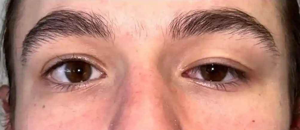
Complications Common to Ptosis Surgery
Overcorrection or undercorrection of lid height is the most common reason for revision surgery across all techniques, and achieving symmetric, appropriately positioned lids, particularly in bilateral cases, requires careful intraoperative assessment and sometimes staged adjustment.
Lagophthalmos, incomplete lid closure, particularly with frontalis sling or more aggressive levator advancement, can predispose to exposure keratopathy, discussed in its own dedicated article on this site, and warrants appropriate lubrication and monitoring postoperatively.
Lid contour irregularity, asymmetric lid crease formation, and, less commonly, more significant complications round out the general risk profile shared across these related but mechanically distinct procedures.
Setting realistic expectations before surgery, including the real possibility of needing a minor adjustment procedure to fine-tune symmetry afterward, generally serves patients better than presenting any single operation as guaranteed to achieve a perfect result on the first attempt.


Document what you see
Two smartphone imaging tools built for everyday clinic use — one for the slit lamp, one for the fundus.
From Choroida — the team behind this siteReferences
- American Academy of Ophthalmology. Basic and Clinical Science Course, Section 7: Oculofacial Plastic and Orbital Surgery.
- Putterman AM, Urist MJ. Müller muscle-conjunctiva resection: technique for treatment of blepharoptosis. Archives of Ophthalmology.
- Bowling B. Kanski’s Clinical Ophthalmology.
Test yourself
A few questions straight from this article.
-
How is levator function measured when planning ptosis surgery?
Levator function is the total excursion of the upper lid between extreme downgaze and extreme upgaze while the frontalis is held still, reflecting how much the levator can still do. -
What does poor levator function favour in the choice of ptosis technique?
When the levator contributes little useful lifting force, techniques that recruit a different muscle, most commonly the frontalis, to do the work of lifting are favoured. -
Which test result is required before Müller's muscle-conjunctival resection is chosen?
Topical phenylephrine temporarily stimulates Müller's muscle and previews how much lift the surgery will achieve, so a demonstrated positive response is critical to selecting this technique. -
What surgical approach does Müller's muscle-conjunctival resection use?
The procedure resects Müller's muscle and overlying conjunctiva from a posterior, internal approach with no external skin incision, making it comparatively less invasive. -
Which underlying mechanism is typically corrected by levator advancement or resection?
Levator advancement or resection is the most commonly performed technique overall, used for typical aponeurotic ptosis where the aponeurosis has stretched or dehisced with age. -
What advantage does the external skin-crease incision give in levator surgery?
Working through an external skin-crease incision allows direct visualisation and precise intraoperative adjustment of both lid height and contour during the procedure. -
For which clinical picture is a frontalis sling reserved?
The frontalis sling is reserved for severe ptosis with poor levator function, most classically congenital ptosis where the levator is poorly developed or non-functioning. -
Which materials are used for the sling in frontalis suspension surgery?
The sling is made of either autologous fascia lata or a synthetic material, connecting the eyelid to the frontalis muscle above the eyebrow so that brow elevation lifts the lid. -
What is the most common reason for revision surgery after ptosis repair?
Over- or undercorrection of lid height is the commonest reason for revision across all ptosis techniques, and achieving symmetry in bilateral cases may need staged adjustment. -
What complication can incomplete lid closure after ptosis surgery predispose to?
Lagophthalmos, seen particularly after frontalis sling or more aggressive levator advancement, can predispose to exposure keratopathy and warrants lubrication and monitoring.
