
Corneal graft rejection is a race against time in the most literal sense, since the endothelial cells under immunologic attack cannot regenerate once lost, and the entire difference between a graft that recovers and one that fails often comes down to how quickly a patient recognizes the warning signs and gets to urgent, aggressive treatment.
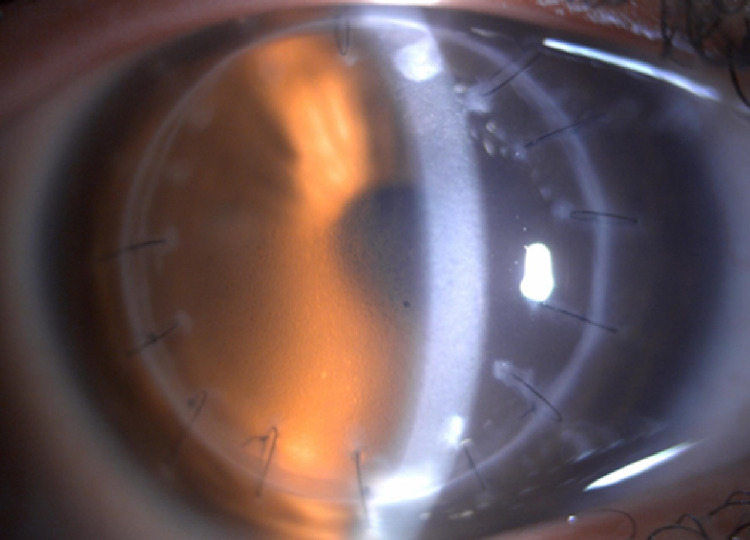
Why Endothelial Rejection Is the One That Matters Most
Corneal grafts can experience rejection affecting different layers, epithelial, subepithelial/stromal, or endothelial, but endothelial rejection is by far the most visually threatening, since the endothelium has no meaningful capacity for cell regeneration, meaning cells lost during a rejection episode are permanently lost, and sufficient endothelial cell loss leads directly to irreversible graft failure and corneal edema.
The Classic RSVP Warning Signs
Patients are typically counseled to watch for and immediately report the following symptoms, often summarized with the mnemonic RSVP, since prompt recognition and treatment meaningfully improves the chance of reversing an episode before it progresses to graft failure:
- Redness
- Sensitivity to light (photophobia)
- Vision decrease
- Pain
Any of these symptoms developing in a corneal transplant recipient, even in isolation, warrants urgent ophthalmic evaluation rather than a wait-and-see approach, given how time-sensitive effective treatment of endothelial rejection is.
All-fit Slit-Lamp Adapter
Record and share exactly what you see at the slit lamp. One adapter fits any slit lamp or surgical microscope — and any smartphone.
From Choroida — the team behind this siteClinical Signs of Endothelial Rejection
- A Khodadoust line, a linear, often curved line of inflammatory cells migrating across the donor endothelium, representing the advancing front of the immune-mediated rejection process and considered the classic, most specific sign of endothelial rejection
- Keratic precipitates on the donor endothelium
- Localized or diffuse corneal edema, corresponding to the area of endothelial cell damage and dysfunction from the rejection process
- Anterior chamber inflammatory reaction, reflecting the broader immune activity
Other Forms of Rejection
- Epithelial rejection presents with an elevated, sometimes faintly visible line of rejected epithelial cells, generally causing less severe visual consequences than endothelial rejection given the epithelium’s capacity to regenerate
- Subepithelial (stromal) rejection produces focal subepithelial infiltrates, also generally less visually threatening than endothelial involvement given the potential for at least partial stromal recovery
Risk Factors for Rejection
- Increased corneal vascularization of the host bed before or after transplantation, providing more direct access for the host immune system to the donor tissue
- Larger graft size and grafts positioned closer to the limbus
- A history of previous graft rejection or failure
- Active ocular surface inflammation at the time of or after transplantation
- Reduced or inconsistent use of prescribed maintenance topical corticosteroids after transplantation
Management
Urgent High-Dose Corticosteroid Treatment
Once endothelial rejection is diagnosed, prompt initiation of intensive topical corticosteroid treatment, often starting with very frequent dosing, is the central intervention, aiming to halt the immune-mediated attack on the endothelium before irreversible cell loss accumulates.
- Periocular or systemic corticosteroids may be added for more severe rejection episodes, given the urgency of controlling the process quickly
- Close, short-interval follow-up during the acute treatment phase allows adjustment of treatment intensity based on the observed response
Maintenance Therapy to Prevent Future Episodes
After an acute episode resolves, ongoing maintenance topical corticosteroid therapy, often at a lower chronic dose, is continued long-term, since corneal grafts, unlike many other transplanted tissues, require sustained topical immunosuppression indefinitely to minimize ongoing rejection risk, given the cornea’s unique relationship with the host immune system.
Patient Education
Because the difference between a reversed rejection episode and graft failure often comes down to how quickly treatment begins, thorough patient education about the RSVP warning signs, provided both after initial transplantation and reinforced at follow-up visits, is a genuinely consequential part of long-term graft care.
A simple message for patients
Teach patients the RSVP warning signs: redness, sensitivity to light, vision decrease, and pain. A graft recipient who has any of these should be seen the same day, even years after surgery, because early treatment with frequent topical steroids and, in some cases, a short course of systemic steroids can reverse an episode. Delay reduces the chance of saving the graft.
What the examiner looks for
Look for a line of keratic precipitates on the endothelium (the Khodadoust line), subepithelial infiltrates, stromal edema, and cells in the chamber. Compare with the findings at the last visit, and check the pressure. Document the graft clarity and the thickness to judge the response.
Prognosis
Endothelial rejection episodes caught early and treated promptly and aggressively have a reasonable chance of reversal, with the graft recovering adequate endothelial function to remain clear.
Delayed recognition or treatment allows progressive, irreversible endothelial cell loss, which can tip a graft into decompensation and eventual failure, underscoring why patient education about these specific warning signs is considered as important to long-term graft survival as the surgical technique and postoperative medication regimen themselves.


Document what you see
Two smartphone imaging tools built for everyday clinic use — one for the slit lamp, one for the fundus.
From Choroida — the team behind this siteReferences
- Panda A, Vanathi M, Kumar A, Dash Y, Priya S. Corneal graft rejection. Surv Ophthalmol. 2007;52:375-396.
- Williams KA, Coster DJ. The immunobiology of corneal transplantation. Transplantation. 2007;84:806-813.
- Khodadoust AA. The allograft rejection reaction: the leading cause of late failure of clinical corneal grafts. Ciba Found Symp. 1973;15:151-167.
- Price MO, Thompson RW Jr, Price FW Jr. Risk factors for various causes of failure in initial corneal grafts. Arch Ophthalmol. 2003;121:1087-1092.
