
Electrooculography, or EOG, measures the standing electrical potential across the eye that arises from the resting activity of the retinal pigment epithelium, providing a functional test of RPE health that is genuinely distinct from what electroretinography measures.
While electroretinography assesses the photoreceptor and inner retinal response to a light flash, EOG instead measures a slower, baseline electrical signal that depends on RPE metabolic function, making the two tests complementary rather than interchangeable despite both being broadly classified as electrophysiologic studies.
Understanding this distinction is essential to understanding why EOG remains the specific, defining test for one particular disease, Best vitelliform macular dystrophy, discussed in relation to vitelliform macular dystrophy in its own dedicated article on this site, even though it has a much narrower range of everyday clinical use than ERG.
Unlike ERG, EOG is performed only in a comparatively small number of specialized electrophysiology laboratories, reflecting its narrow indication rather than any particular technical difficulty in performing the test itself.
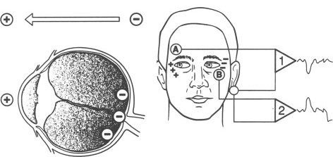
What EOG Actually Measures
The retina maintains a standing electrical potential between the cornea, relatively positive, and the back of the eye, relatively negative, generated largely by the resting membrane potential of the retinal pigment epithelium.
This potential changes measurably between dark and light adaptation, and the specific pattern of that change, rather than the raw potential itself, is what the test actually quantifies and reports.
Because the signal depends on RPE function specifically, EOG is most useful for diseases primarily affecting the RPE, rather than diseases affecting photoreceptors or inner retinal cells, which is the underlying reason its clinical utility is concentrated in one particular category of retinal disease.
The Arden Ratio
The test is performed by having the patient make repeated horizontal eye movements between two fixed points while electrodes near the eyes record the changing potential, first in the dark and then after exposure to light.
The light peak, the maximum potential recorded after light exposure, is compared to the dark trough, the minimum potential recorded during dark adaptation, and this ratio, called the Arden ratio, is the key quantitative result reported from the test.
A normal Arden ratio reflects healthy RPE function responding appropriately to the light-dark transition, while an abnormally low ratio indicates a defect in this RPE-dependent light response, regardless of how the overlying photoreceptors and retina appear structurally on exam or imaging.
Fundus Explorer Pro
Photograph the retinal findings described here with the phone already in your pocket — 22 D optics and built-in illumination in one handheld unit.
From Choroida — the team behind this siteWhy EOG Is the Defining Test for Best Disease
Best vitelliform macular dystrophy is characterized by a severely reduced Arden ratio that is disproportionate to, and often precedes, any visible fundus abnormality, reflecting a generalized RPE dysfunction extending well beyond the visible macular lesion itself.
This severely abnormal EOG, even in family members with a normal-appearing macula who have not yet developed the classic “egg yolk” lesion, is a hallmark diagnostic and genetic-carrier-screening finding specific to this condition.
In most other macular and retinal dystrophies, the EOG is normal or only mildly abnormal, which is why a severely reduced Arden ratio carries such specific diagnostic weight when Best disease is being considered in the differential.
Genetic testing for the causative BEST1 gene mutation has become increasingly available alongside EOG, and the two tests are complementary, with genetic confirmation adding certainty in cases where the clinical and electrophysiologic picture leaves some ambiguity.
Clinical Uses
- Confirming a diagnosis of Best vitelliform macular dystrophy, particularly in atypical or early presentations where the classic lesion is not yet fully developed
- Screening asymptomatic family members of a patient with confirmed Best disease, since the EOG can be abnormal even before any visible fundus change appears
- Distinguishing Best disease from adult-onset vitelliform maculopathy, discussed in its own dedicated article on this site, which typically shows a normal or only mildly reduced Arden ratio despite a visually similar-appearing macular lesion
Limitations
EOG requires sustained patient cooperation over a relatively long testing session, including repeated eye movements during both dark and light adaptation phases, making it more demanding for young children or poorly cooperative patients than a standard ERG.
The test’s clinical utility is genuinely narrow compared to ERG, since most retinal and macular conditions do not show a specific or diagnostically useful EOG abnormality, which is why it is ordered selectively rather than as a routine part of a broad electrophysiology workup.
For this reason, EOG is best thought of not as a general-purpose retinal function test but as a targeted confirmatory tool reserved for the specific clinical question of suspected Best disease or a closely related bestrophinopathy.


Document what you see
Two smartphone imaging tools built for everyday clinic use — one for the slit lamp, one for the fundus.
From Choroida — the team behind this siteReferences
- American Academy of Ophthalmology. Basic and Clinical Science Course, Section 12: Retina and Vitreous.
- Marmor MF, Zrenner E. Standard for clinical electro-oculography. International Society for Clinical Electrophysiology of Vision. Archives of Ophthalmology.
- Constable PA, Bach M, Frishman LJ, et al. ISCEV standard for clinical electro-oculography. Documenta Ophthalmologica.
Test yourself
A few questions straight from this article.
-
Electrooculography provides a functional test of which retinal structure?
EOG measures the standing electrical potential arising from the resting activity of the retinal pigment epithelium, making it a functional test of RPE health. -
How does electroretinography differ from electrooculography in what it assesses?
Electroretinography assesses the photoreceptor and inner retinal response to a light flash, while EOG measures a slower baseline signal dependent on RPE metabolic function. -
In the standing potential of the eye, which polarity relationship holds?
The retina maintains a standing potential with the cornea relatively positive and the back of the eye relatively negative, generated largely by the RPE resting membrane potential. -
What is the patient asked to do during an electrooculography recording?
The patient makes repeated horizontal eye movements between two fixed points while electrodes near the eyes record the changing potential, first in the dark and then after light exposure. -
The Arden ratio compares which two measurements?
The light peak, the maximum potential after light exposure, is compared with the dark trough, the minimum during dark adaptation, and this ratio is the key reported result. -
An abnormally low Arden ratio indicates which abnormality?
An abnormally low ratio indicates a defect in the RPE-dependent light response, regardless of how the overlying photoreceptors and retina appear on examination or imaging. -
Which electrooculography finding characterises Best vitelliform macular dystrophy?
Best disease shows a severely reduced Arden ratio disproportionate to, and often preceding, any visible fundus abnormality, reflecting generalised RPE dysfunction beyond the macular lesion. -
Why is electrooculography useful in asymptomatic relatives of a patient with Best disease?
The EOG can be severely abnormal in family members whose macula looks normal and who have not yet developed the classic egg yolk lesion, making it valuable for carrier screening. -
Which condition shows a normal or only mildly reduced Arden ratio despite a similar macular lesion?
Adult-onset vitelliform maculopathy typically shows a normal or only mildly reduced Arden ratio despite a macular lesion that can look visually similar to that of Best disease. -
Which limitation makes electrooculography difficult in young or poorly cooperative patients?
EOG requires sustained cooperation over a relatively long session, with repeated eye movements during both dark and light adaptation, making it more demanding than a standard ERG.

Writes and reviews the clinical articles on Educate. Every post is checked against peer-reviewed sources before it goes live.


