
Anterior lenticonus is a conical or globular protrusion of the central anterior lens surface, and its strong, well-established association with Alport syndrome means that recognizing this specific lens shape at the slit lamp can be the finding that leads to identification of a serious hereditary kidney disease in a patient, or their family, who did not yet carry that diagnosis.
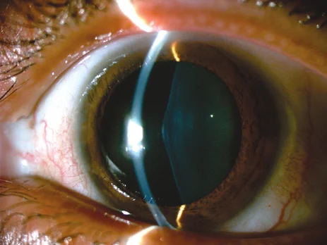
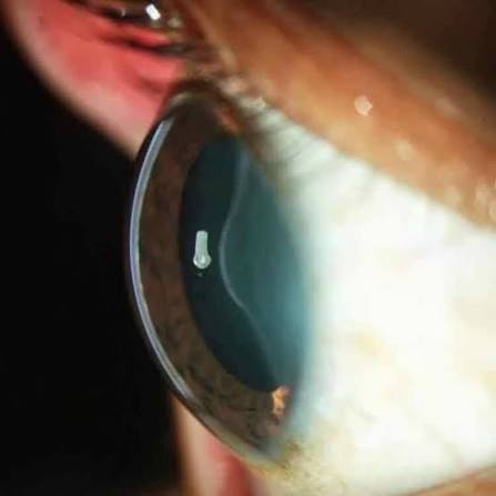
What Anterior Lenticonus Looks Like
Rather than the normal smooth, gently curved anterior lens surface, anterior lenticonus produces a localized, cone-shaped or dome-shaped bulge at the central anterior lens capsule and cortex, thought to result from a weakness or thinning of the central anterior lens capsule that allows the underlying lens cortex to bulge forward under normal intraocular pressure.
The Oil-Droplet Sign
One of the most characteristic and diagnostically useful findings in anterior lenticonus is the “oil droplet” sign seen on retinoscopy or, similarly, on direct ophthalmoscopy: because the conical protrusion has different optical power than the surrounding lens, it produces a distinctive dark, round, oil-droplet-like reflex against the surrounding red reflex, a finding that is often more readily appreciated on retinoscopy than on slit-lamp examination alone, and one every clinician evaluating a patient with known or suspected Alport syndrome should specifically look for.
All-fit Slit-Lamp Adapter
Record and share exactly what you see at the slit lamp. One adapter fits any slit lamp or surgical microscope — and any smartphone.
From Choroida — the team behind this siteAssociation With Alport Syndrome
Alport syndrome is a hereditary condition, most commonly X-linked, caused by mutations in genes encoding type IV collagen, including COL4A5, affecting the basement membranes of the kidney, inner ear, and eye, and anterior lenticonus is one of the most specific and recognized ocular manifestations of this syndrome.
- Progressive hereditary nephritis, often progressing to end-stage renal disease, is the dominant and most clinically significant feature of Alport syndrome
- Sensorineural hearing loss, typically progressive, is another core systemic feature
- Anterior lenticonus occurs in a meaningful proportion of affected males with the X-linked form and is considered one of the more specific ocular findings for this diagnosis, since it is uncommon outside the context of Alport syndrome
Other Ocular Findings in Alport Syndrome
- A perimacular fleck retinopathy, with yellow or white flecks in a ring pattern around the macula, another recognized and relatively specific ocular finding
- Posterior polymorphous corneal dystrophy has also been reported in association with Alport syndrome in some patients (see posterior polymorphous corneal dystrophy)
- Recurrent corneal erosion has been described in some affected individuals as well
Clinical and Diagnostic Approach
- Retinoscopy or direct ophthalmoscopy specifically looking for the oil-droplet reflex in any patient with known or suspected Alport syndrome, or in any young patient found to have anterior lenticonus for another reason, since this finding should itself prompt consideration of the diagnosis if not already established
- A careful family and personal history for hereditary nephritis and sensorineural hearing loss
- Coordination with nephrology for renal function assessment and genetic testing when Alport syndrome is suspected based on ocular findings in a patient not previously known to have the condition
- Dilated fundus examination looking for the characteristic perimacular flecks, providing additional supportive evidence
Management
Managing the Lens Finding
Anterior lenticonus itself is managed based on its visual impact: mild cases without significant refractive or optical disturbance may simply be observed, while more pronounced lenticonus causing irregular astigmatism or significant visual impairment can be managed with cataract extraction and intraocular lens implantation, which is generally effective at restoring good vision once the abnormal lens tissue is removed.
Coordinated Systemic Care
Given the seriousness of the renal component of Alport syndrome, identification of anterior lenticonus in a patient not previously known to have the condition should prompt prompt referral for nephrology evaluation, since early recognition and management of progressive nephritis has real implications for long-term kidney health and transplant planning.
Prognosis
Visual outcomes after surgical management of anterior lenticonus, when needed, are generally very good, since removing the abnormally shaped lens tissue and replacing it with a standard intraocular lens addresses the optical problem directly.
The greater long-term significance of recognizing anterior lenticonus often lies not in the eye itself but in the systemic diagnosis it can point toward, underscoring why this specific lens finding deserves more than a passing note in the chart when identified.


Document what you see
Two smartphone imaging tools built for everyday clinic use — one for the slit lamp, one for the fundus.
From Choroida — the team behind this siteReferences
- Kato T, Watanabe Y, Nakayasu K, Kanai A, Yajima Y. The ultrastructure of the lens capsule abnormalities in Alport’s syndrome. Jpn J Ophthalmol. 1998;42:401-405.
- Colville DJ, Savige J. Alport syndrome: a review of the ocular manifestations. Ophthalmic Genet. 1997;18:161-173.
- Kashtan CE. Alport syndrome: an inherited disorder of renal, ocular, and cochlear basement membranes. Medicine (Baltimore). 1999;78:338-360.
- Kato T, Watanabe Y, Nakayasu K, Kanai A, Yajima Y. Anterior lenticonus and Alport syndrome. Br J Ophthalmol. 1998;82:1210.
