
Vitreous hemorrhage is bleeding into the vitreous cavity, and it presents a genuinely distinctive diagnostic problem: the blood itself obscures the very structures a clinician needs to examine to find out why it is there in the first place.
The clinical approach has to work around this blind spot, using history, the pattern of visual loss, and imaging that does not depend on a clear view of the fundus, until the haemorrhage clears enough to actually see what caused it.
Causes
- Proliferative diabetic retinopathy – the most common cause in most populations, from bleeding of fragile neovascular vessels
- Posterior vitreous detachment with an associated retinal tear, where a torn retinal vessel bleeds into the vitreous cavity
- Retinal vein occlusion with secondary neovascularisation, following the same mechanism as diabetic disease but with a different underlying vascular insult
- Ocular trauma, blunt or penetrating, a common cause particularly in younger patients
- Retinal macroaneurysm rupture, less common but a recognised cause of sudden, sometimes dramatic haemorrhage
- Wet age-related macular degeneration, when a neovascular membrane bleeds significantly beyond the subretinal space
- Rarer causes including Terson syndrome (vitreous haemorrhage associated with subarachnoid haemorrhage), sickle cell retinopathy, and blood dyscrasias
Age is a genuinely useful first filter: diabetic retinopathy and vein occlusion dominate in older patients, while trauma and posterior vitreous detachment with a retinal tear are more prominent considerations in younger patients presenting with sudden vitreous haemorrhage.
Clinical Presentation
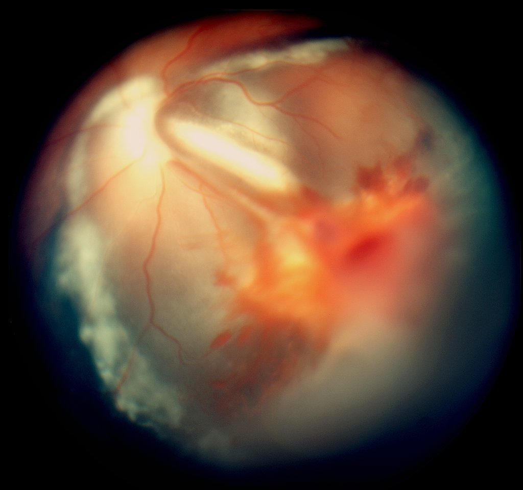
Patients typically describe sudden onset of floaters, often numerous and described as cobwebs, soot, or a shower of dark spots, sometimes progressing rapidly to significant visual blurring or loss depending on the volume of blood.
Small haemorrhages may cause only mild floaters with relatively preserved vision, while dense haemorrhage can reduce vision to hand motions or light perception, with the fundus view completely obscured on examination.
Pain is not typically a feature unless the haemorrhage is associated with significantly raised intraocular pressure or an underlying condition, such as trauma, that independently causes pain.
Fundus Explorer Pro
Photograph the retinal findings described here with the phone already in your pocket — 22 D optics and built-in illumination in one handheld unit.
From Choroida — the team behind this siteDiagnostic Evaluation
Dilated examination is attempted first, since even a partial view can sometimes identify the source, but when the view is too poor, B-scan ultrasonography becomes the key investigation, assessing for retinal detachment, a mass lesion, or other structural abnormality that plain examination cannot reveal through dense blood.
A history of diabetes, prior retinal vein occlusion, trauma, or systemic conditions associated with bleeding should be specifically sought, since this history often narrows the differential substantially even before the fundus can be visualised directly.
Serial examination as the haemorrhage clears, sometimes over days to weeks, eventually allows direct visualisation of the underlying cause, and this staged approach, imaging first and confirmatory examination once the view improves, is standard rather than a failure to reach an immediate diagnosis.
Differential Diagnosis of Sudden Floaters and Vision Loss
- Posterior vitreous detachment without haemorrhage, generally with fewer, more discrete floaters and preserved vision
- Retinal detachment, which can coexist with vitreous haemorrhage or present independently with a visual field defect
- Endophthalmitis, distinguished by pain, redness, and a relevant history of recent surgery or trauma
- Central retinal artery or vein occlusion, generally with a more profound, sudden visual change and a different fundus appearance once visible
Management
Observation
Many cases of vitreous haemorrhage, particularly smaller volumes without evidence of retinal detachment on B-scan, are managed with observation, allowing time for spontaneous clearing while addressing any identifiable underlying cause, such as optimising diabetic control or completing panretinal photocoagulation once the view allows.
Treating the Underlying Cause
Panretinal photocoagulation is completed or intensified once the view permits in diabetic or ischaemic vein occlusion-related haemorrhage, aiming to regress the neovascularisation responsible for bleeding and reduce the risk of recurrence.
Anti-VEGF therapy can be used to reduce neovascular activity and may help clear haemorrhage in some cases, often used alongside or as a bridge to definitive laser treatment.
Surgery
Pars plana vitrectomy is indicated for dense, non-clearing haemorrhage, particularly when B-scan raises concern for retinal detachment, or when the haemorrhage is preventing necessary treatment such as panretinal photocoagulation from being delivered. Vitrectomy clears the blood directly and allows any underlying retinal pathology to be addressed at the same time.
Prognosis
Visual recovery depends heavily on the underlying cause rather than the haemorrhage itself; haemorrhage from a benign posterior vitreous detachment without a retinal tear generally has an excellent prognosis once it clears, while haemorrhage associated with retinal detachment or advanced proliferative disease carries a more guarded outlook.
Recurrent haemorrhage is common in diabetic retinopathy and vein occlusion until the underlying neovascular drive is adequately treated, which is why definitive treatment of the cause, not just clearance of the current episode, is the actual goal of management.
Prompt B-scan imaging when the view is poor, rather than simply waiting for the blood to clear before further assessment, is what allows retinal detachment to be identified and treated early, and this single step is often what separates a good visual outcome from a preventable one.


Document what you see
Two smartphone imaging tools built for everyday clinic use — one for the slit lamp, one for the fundus.
From Choroida — the team behind this siteReferences
- Spraul CW, Grossniklaus HE. Vitreous hemorrhage. Survey of Ophthalmology. 1997.
- Lindgren G, Sjodell L, Lindblom B. A prospective study of dense spontaneous vitreous hemorrhage. American Journal of Ophthalmology. 1995.
- Wiznia RA. Posterior vitreous detachment and asteroid hyalosis. American Journal of Ophthalmology. 1987.
- Vitreous Hemorrhage. EyeWiki, American Academy of Ophthalmology.
- Vitreous Hemorrhage. StatPearls, NCBI Bookshelf.
Test yourself
A few questions straight from this article.
-
What makes vitreous haemorrhage a distinctive diagnostic problem?
Blood in the vitreous cavity obscures the very fundus structures a clinician must examine to establish why the bleeding happened, so the approach must work around that blind spot. -
What is the most common cause of vitreous haemorrhage in most populations?
Bleeding from fragile neovascular vessels in proliferative diabetic retinopathy is the leading cause of vitreous haemorrhage in most populations. -
How does posterior vitreous detachment produce a vitreous haemorrhage?
When posterior vitreous detachment creates a retinal tear, a torn retinal vessel can bleed into the vitreous cavity. -
Which causes are most prominent in a younger patient with sudden vitreous haemorrhage?
Age is a useful first filter: diabetic retinopathy and vein occlusion dominate in older patients, while trauma and posterior vitreous detachment with a retinal tear lead in younger ones. -
How do patients typically describe the onset of vitreous haemorrhage?
Patients describe a sudden shower of dark spots, cobwebs or soot, sometimes progressing quickly to blurring or marked visual loss depending on the volume of blood. -
Which investigation is key when dense vitreous haemorrhage prevents any fundus view?
B-scan ultrasonography assesses for retinal detachment, a mass lesion or other structural abnormality that examination cannot reveal through dense blood. -
How does posterior vitreous detachment without haemorrhage usually differ at presentation?
Uncomplicated posterior vitreous detachment generally causes fewer, more discrete floaters and vision is preserved, unlike the dense shower and visual loss of vitreous haemorrhage. -
What is the role of panretinal photocoagulation after vitreous haemorrhage from proliferative disease?
Laser is completed or intensified as soon as the view allows, aiming to regress the neovascularisation responsible for the bleeding and reduce the risk of recurrence. -
What is the main indication for pars plana vitrectomy in vitreous haemorrhage?
Vitrectomy is indicated for dense, non-clearing blood, particularly when B-scan raises concern for retinal detachment or the haemorrhage is blocking necessary laser treatment. -
What most determines visual recovery after a vitreous haemorrhage?
Bleeding from a benign posterior vitreous detachment without a tear recovers excellently, while haemorrhage with retinal detachment or advanced proliferative disease carries a far more guarded outlook.

Writes and reviews the clinical articles on Educate. Every post is checked against peer-reviewed sources before it goes live.


