
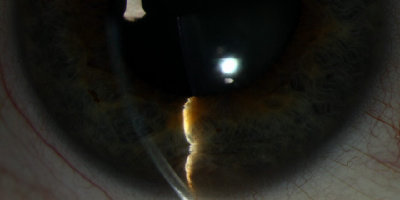
An iris cyst can look, at first glance, remarkably similar to a solid iris tumor, both presenting as a discrete elevation or mass on or behind the iris, but the distinction matters enormously for management, since a cyst is typically observed while a solid melanocytic lesion may need active surveillance or intervention, which is exactly why the specific tools that reveal a lesion’s fluid-filled nature, transillumination and ultrasound biomicroscopy, are central to correct evaluation.
Primary Iris Cysts
Iris Pigment Epithelial Cysts
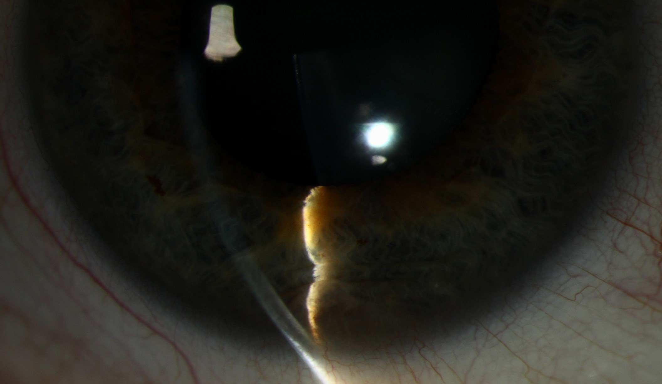
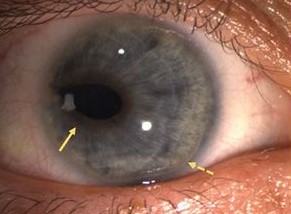
These arise from the posterior pigment epithelial layer of the iris and are the most common type of primary iris cyst, typically located at the pupillary margin, mid-zone, or peripheral iris, and often discovered incidentally, since they frequently cause no symptoms and remain stable indefinitely.
Iris Stromal Cysts
Less common than pigment epithelial cysts, these arise within the anterior iris stroma itself, tend to present earlier in life, sometimes congenitally or in early childhood, and are more likely to be clinically significant, since they can enlarge, distort the pupil, or in some cases affect vision or intraocular pressure through mechanical angle crowding.
Secondary Iris Cysts
- Medication-induced cysts, associated with certain topical medications, notably miotic agents used historically for glaucoma or accommodative esotropia treatment, and some prostaglandin analogues in rare reported cases
- Implantation cysts, arising after surgical or traumatic introduction of surface epithelium into the anterior chamber, where it can proliferate and form a cystic structure, sometimes growing progressively and causing significant complications if not addressed
- Parasitic cysts, a rare cause in endemic regions
All-fit Slit-Lamp Adapter
Record and share exactly what you see at the slit lamp. One adapter fits any slit lamp or surgical microscope — and any smartphone.
From Choroida — the team behind this siteClinical Features
- A translucent to variably pigmented, dome-shaped elevation, which can arise from the iris pigment epithelium (posterior surface), the iris stroma (anterior surface), or occasionally project into the anterior chamber angle
- Generally slow-growing or entirely stable over time for primary cysts, in contrast to the more variable and sometimes progressive behavior of implantation cysts
- Larger cysts can cause mechanical effects, including pupillary distortion, induced astigmatism, or, when extending into the angle, secondary angle-closure glaucoma
Distinguishing Cysts From Solid Tumors
Transillumination
Shining a light through the sclera and observing the lesion from the pupil, a cyst characteristically transilluminates readily, appearing bright and translucent, since it is fluid-filled, while a solid melanocytic tumor blocks transmitted light and appears as a dark shadow, a simple, valuable bedside distinguishing test.
Ultrasound Biomicroscopy
This high-frequency ultrasound technique directly visualizes the lesion’s internal characteristics, showing the classic smooth-walled, echolucent (fluid-filled) appearance of a cyst in contrast to the solid, echogenic internal pattern of a tumor, and is particularly valuable for lesions arising from the posterior iris surface that are not fully visible on direct slit-lamp examination.
Management
Observation
The majority of primary iris pigment epithelial cysts and many iris stromal cysts are managed with observation alone, given their generally benign, stable natural history, with periodic examination to confirm continued stability.
Intervention for Symptomatic or Growing Cysts
- Cysts causing significant visual symptoms, mechanical pupillary distortion, or secondary glaucoma from angle involvement may warrant intervention
- Options include needle aspiration, sometimes combined with intracystic injection of a sclerosing agent, and surgical excision for cysts not adequately managed by aspiration alone, particularly implantation cysts, which often behave more aggressively and are more likely to require definitive surgical removal
Managing Secondary Cysts
Medication-induced cysts often regress with discontinuation of the causative agent when this is feasible, while implantation cysts generally require surgical management given their tendency toward progressive growth if left untreated.
Prognosis
Primary iris cysts generally have an excellent prognosis, with most remaining stable and asymptomatic indefinitely, requiring nothing more than periodic observation.
Implantation cysts and other secondary cysts with a tendency toward progressive growth require closer monitoring and a lower threshold for intervention, but even these generally respond well to appropriate treatment once correctly identified and distinguished from a solid tumor requiring a fundamentally different management approach.


Document what you see
Two smartphone imaging tools built for everyday clinic use — one for the slit lamp, one for the fundus.
From Choroida — the team behind this siteReferences
- Shields JA, Shields CL, Lois N, Mercado G. Iris cysts in children: classification, incidence, and management. Br J Ophthalmol. 1999;83:334-338.
- Shields JA, Kline MW, Augsburger JJ. Primary iris cysts: a review of the literature and report of 62 cases. Br J Ophthalmol. 1984;68:152-166.
- Behrouzi Z, Khodadoust A. Epithelial iris cyst treatment with double freeze-thaw cryotherapy. Ophthalmology. 2003;110:1885-1889.
- Marigo FA, Finger PT, McCormick SA, Iacob CE, Grossniklaus HE, Bekerman VP. Iris and ciliary body melanomas: ultrasound biomicroscopy for the diagnosis and management. Ophthalmology. 2000;107:1929-1936.
