
Spheroidal degeneration, also called climatic droplet keratopathy or Labrador keratopathy, is a slowly progressive corneal change caused by cumulative ultraviolet exposure, most often seen in people who have spent decades working outdoors in sunny, dry, or high-altitude environments, and it is a useful reminder that the cornea, not just the skin and the lens, accumulates measurable damage from a lifetime of sun exposure.
What Causes the Deposits
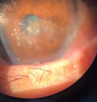
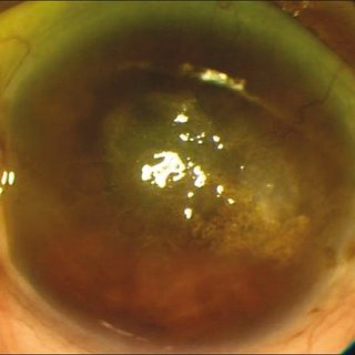
Chronic ultraviolet exposure is believed to damage corneal stromal proteins, leading to the deposition of an abnormal, golden-brown, translucent protein material within the superficial stroma, most concentrated in the interpalpebral zone where UV exposure is greatest, similar in distribution logic to the exposure pattern seen in pterygium and band keratopathy.
Other environmental factors, including chronic dryness, wind, and dust exposure, are thought to contribute alongside ultraviolet light itself, which is why the condition clusters in populations with a combination of high sun exposure and harsh outdoor working conditions.
Clinical Features and Staging
- Early disease: fine, golden-brown, translucent droplets in the superficial stroma, typically most concentrated within the interpalpebral zone, often first appearing nasally and temporally near the limbus
- Progressive disease: droplets coalesce and can extend centrally, sometimes forming larger, more confluent opacities
- Advanced disease: significant central corneal involvement can reduce vision, and the surface can become irregular
- The condition is typically bilateral and progresses very slowly over years to decades, tracking with cumulative sun exposure over a lifetime rather than any single event
All-fit Slit-Lamp Adapter
Record and share exactly what you see at the slit lamp. One adapter fits any slit lamp or surgical microscope — and any smartphone.
From Choroida — the team behind this sitePopulations Affected
Spheroidal degeneration is classically described in populations with prolonged outdoor occupational exposure in bright, often reflective environments, historically including fishermen, particularly in regions such as Labrador where the condition was first well described, as well as outdoor workers in high-altitude, desert, and other high-UV environments worldwide.
Age and cumulative years of outdoor exposure are the strongest risk factors, more so than any single period of intense exposure.
Differential Diagnosis
- Band keratopathy, which has a chalky white, calcific appearance rather than the golden-brown, translucent quality of spheroidal degeneration, and a different underlying cause (see band keratopathy)
- Salzmann nodular degeneration, which is elevated and more discretely nodular rather than the diffuse, droplet-like pattern of spheroidal degeneration (see Salzmann nodular degeneration)
- Lipid keratopathy, usually associated with prior corneal vascularization or inflammation, distinguished by its whiter, more crystalline appearance and clinical context
Evaluation
Diagnosis is generally straightforward from the characteristic clinical appearance and a history of prolonged outdoor occupational or environmental UV exposure.
Slit-lamp examination with careful attention to the interpalpebral zone and comparison of the degree of involvement between the central and peripheral cornea helps stage severity and guide the decision about intervention.
Management
Prevention
UV-protective sunglasses and broad-brimmed hats are the primary preventive measures for those with ongoing significant outdoor exposure, and this counseling is worth giving proactively to patients in high-risk occupations even before significant deposits have developed, since the damage accumulates gradually and is not reversible once established.
Observation
Mild to moderate disease without significant visual impact is generally observed, since the condition progresses very slowly and many patients never develop vision-threatening central involvement.
Surgical Treatment
Visually significant central involvement can be treated with superficial keratectomy, removing the superficial stromal deposits, sometimes combined with phototherapeutic keratectomy to smooth the corneal surface, similar in principle to management of other superficial corneal deposit disorders.
Deeper or more extensive disease not adequately addressed by superficial techniques may rarely require lamellar or penetrating keratoplasty in severe, advanced cases.
Prognosis
Most affected individuals have mild to moderate disease that does not significantly threaten vision over a normal lifetime, particularly with UV protection to limit further progression.
Surgical treatment for the minority with visually significant central involvement is generally effective, though ongoing sun protection remains important afterward to reduce the risk of recurrent deposit formation given that the underlying UV exposure risk, if occupational, often continues.


Document what you see
Two smartphone imaging tools built for everyday clinic use — one for the slit lamp, one for the fundus.
From Choroida — the team behind this siteReferences
- Freedman A. Labrador keratopathy: bilateral, spheroid, degeneration of the cornea. Arch Ophthalmol. 1965;74:198-202.
- Gray RH, Johnson GJ, Freedman A. Climatic droplet keratopathy. Surv Ophthalmol. 1992;36:241-253.
- Norn MS. Spheroid degeneration of the cornea and conjunctiva. Acta Ophthalmol (Copenh). 1979;57:1-13.
- Karp CL, Scott IU, Green WR, Cameron A, Fine SL. Climatic droplet keratopathy: a review. Surv Ophthalmol. 1999;44:104-116.
