
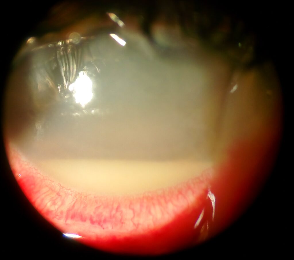
Endogenous and exogenous endophthalmitis are two fundamentally different ways an eye can become infected internally, distinguished by the route organisms take to reach the vitreous cavity, and this single distinction shapes essentially everything about how each is worked up and managed.
Exogenous endophthalmitis, discussed in relation to acute postoperative endophthalmitis in its own dedicated article on this site, results from organisms introduced directly into the eye, most often through surgery or penetrating trauma, while endogenous endophthalmitis, discussed in its own dedicated article on this site, results from organisms reaching the eye through the bloodstream from a distant source of infection elsewhere in the body.
Understanding which pattern a given case represents is not just an academic classification exercise: it determines whether the workup should focus entirely on the eye itself or should extend outward to find and treat a serious systemic infection the eye finding may be the first sign of.
Exogenous cases are considerably more common overall in most general ophthalmic practice, simply given the sheer volume of routine intraocular surgery performed, but endogenous cases carry a distinct urgency of their own given the systemic illness they represent.
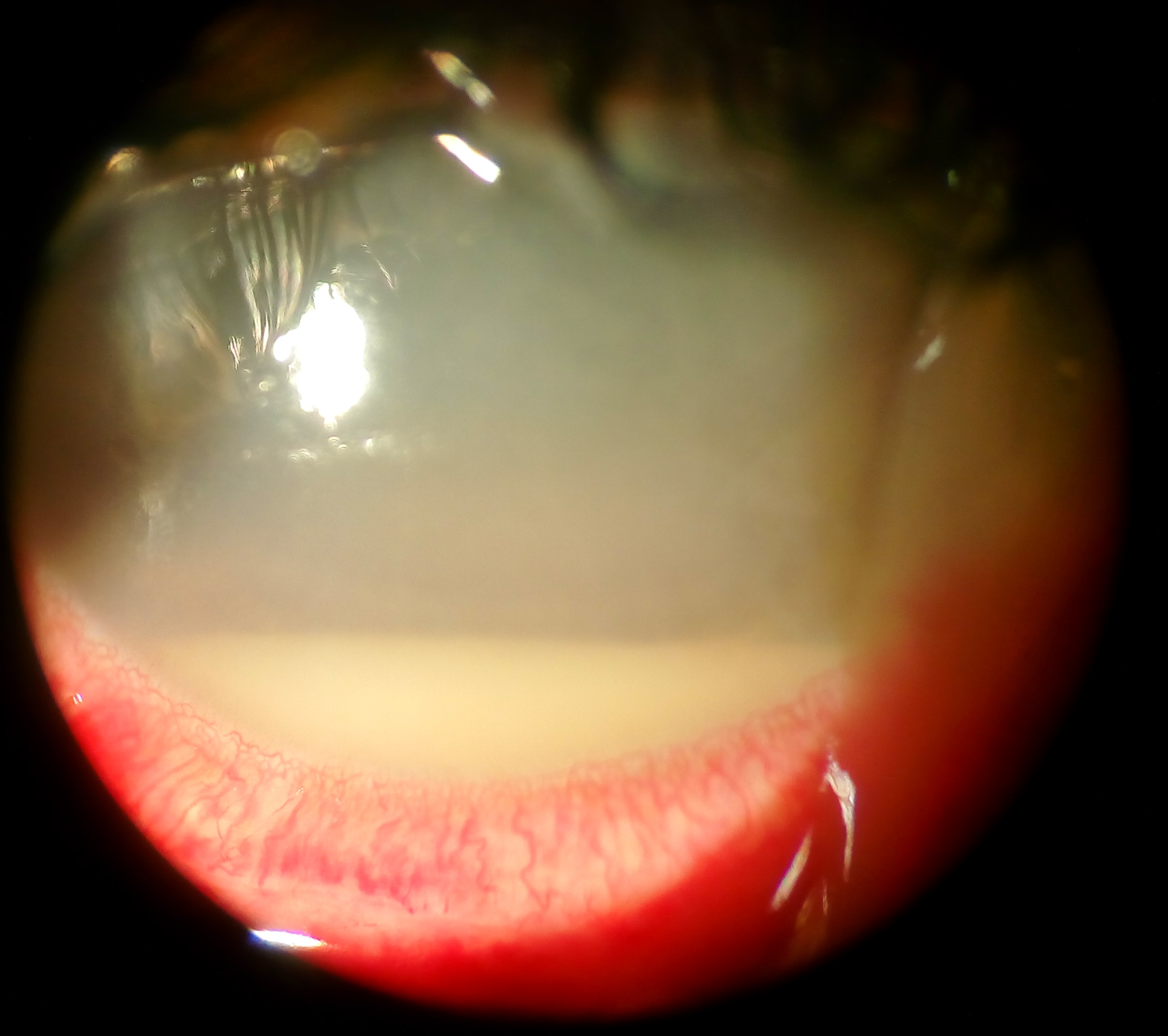
Exogenous Endophthalmitis: The Local Route
Organisms enter the eye directly, most commonly through a surgical incision, particularly after cataract surgery, or through a penetrating injury that breaches the eye’s normal protective barriers.
Because the infection’s origin is entirely local, the workup for exogenous endophthalmitis is correspondingly focused: identifying the causative organism from an aqueous or vitreous sample, and treating with intravitreal antibiotics, without any expectation of finding or needing to search for a separate, distant infectious source.
The timeline is often more predictable in exogenous cases, with acute presentations clustering in the days following a surgical or traumatic breach of the eye’s normal barrier, discussed in relation to acute postoperative endophthalmitis on this site, though the chronic, indolent form discussed in relation to chronic postoperative endophthalmitis can present much later.
Endogenous Endophthalmitis: The Bloodstream Route
Organisms reach the eye through hematogenous spread from a distant infectious focus, most commonly a source such as endocarditis, an abscess, an indwelling catheter-related bloodstream infection, or a urinary tract source in an at-risk patient.
Because the eye is essentially a secondary site of infection in this pattern, endogenous endophthalmitis is often the presenting sign that prompts discovery of a serious, sometimes previously unrecognized, systemic infection, making the ophthalmologist’s finding a genuine trigger for a much broader medical workup.
Risk factors for endogenous endophthalmitis, including intravenous drug use, immunosuppression, indwelling catheters, and recent hospitalization or invasive procedures, reflect the systemic illness and vulnerability that predisposes to bloodstream seeding in the first place.
Fundus Explorer Pro
Photograph the retinal findings described here with the phone already in your pocket — 22 D optics and built-in illumination in one handheld unit.
From Choroida — the team behind this siteWhy the Distinction Drives Different Workups
- Exogenous endophthalmitis: workup centers on the eye itself, with intraocular sampling for culture and prompt intravitreal treatment, and no expectation of a broader systemic source needing investigation
- Endogenous endophthalmitis: workup extends well beyond the eye, including blood cultures, echocardiography when endocarditis is suspected, and imaging to search for an occult abscess or other infectious focus
- Systemic antimicrobial therapy is typically essential in endogenous cases, treating the underlying bloodstream infection alongside local intraocular treatment, whereas exogenous cases are usually managed with local intraocular treatment as the primary intervention
- Coordination with infectious disease and other relevant specialists is far more central to managing endogenous cases, given the systemic infection driving the ocular finding
Overlapping Clinical Features
Both forms can present with pain, redness, decreased vision, and vitritis, and the acute clinical picture at the eye itself does not always immediately distinguish which pattern is present without a careful history specifically probing for a recent surgical or traumatic event versus systemic symptoms and risk factors for bloodstream infection.
A history of recent eye surgery or trauma points strongly toward exogenous infection, while fever, systemic illness, or known risk factors for bacteremia or fungemia point toward an endogenous source, and asking these specific, targeted history questions is often what actually separates the two clinically.
Management Principles
Prompt intraocular sampling and intravitreal antibiotic or antifungal therapy are central to both forms, following broadly similar local treatment principles once the diagnosis of endophthalmitis of either type is confirmed.
Systemic treatment, additional systemic workup, and management of the underlying infectious source are specifically required for endogenous cases, reflecting the genuinely different, broader scope of disease that endogenous endophthalmitis represents compared to a purely local, exogenous infection.
An ophthalmologist recognizing endogenous endophthalmitis is, in effect, often the first clinician to identify a serious, potentially life-threatening systemic infection, which is a responsibility worth taking seriously well beyond the immediate ocular treatment itself.


Document what you see
Two smartphone imaging tools built for everyday clinic use — one for the slit lamp, one for the fundus.
From Choroida — the team behind this siteReferences
- Jackson TL, Eykyn SJ, Graham EM, Stanford MR. Endogenous bacterial endophthalmitis: a 17-year prospective series and review of 267 reported cases. Survey of Ophthalmology.
- American Academy of Ophthalmology. Basic and Clinical Science Course, Section 9: Uveitis and Ocular Inflammation.
- Durand ML. Endophthalmitis. Clinical Microbiology and Infection.
Test yourself
A few questions straight from this article.
-
What single feature distinguishes endogenous from exogenous endophthalmitis?
The two forms are separated by how organisms arrive inside the eye, and that single distinction shapes the whole workup and management of each. -
What is the expected scope of the infectious workup in exogenous endophthalmitis?
Because the origin is entirely local, the workup is confined to identifying the organism from an aqueous or vitreous sample and treating intravitreally. -
How do organisms reach the eye in endogenous endophthalmitis?
In the endogenous pattern the eye is a secondary site, seeded through the bloodstream from an infection elsewhere in the body. -
Which form of endophthalmitis is more common in general ophthalmic practice, and why?
Exogenous cases predominate simply because of how much intraocular surgery is performed, although endogenous cases carry their own urgency from the systemic illness behind them. -
Which investigations belong to the systemic workup of endogenous endophthalmitis?
The search extends well beyond the eye: blood cultures, echocardiography when endocarditis is suspected, and imaging to find an occult abscess or other focus. -
Which treatment element is specifically required in endogenous endophthalmitis?
Endogenous disease needs systemic treatment of the underlying infection alongside local intraocular therapy, whereas exogenous cases are managed mainly with local treatment. -
Which clinical features are shared by endogenous and exogenous endophthalmitis?
Both forms can look identical at the eye itself, so the ocular picture alone often fails to separate them without a targeted history. -
Which history finding points toward an endogenous rather than exogenous source of endophthalmitis?
Systemic illness and bloodstream-infection risk factors point to an endogenous source, while recent surgery or trauma points strongly to an exogenous one. -
Which of these is a recognised distant focus seeding the eye in endogenous endophthalmitis?
Typical sources include endocarditis, an abscess, an indwelling catheter-related bloodstream infection, and a urinary tract source in an at-risk patient. -
Which patient factor increases the risk of endogenous endophthalmitis?
Risk factors such as intravenous drug use, immunosuppression, indwelling catheters and recent hospitalisation all reflect vulnerability to bloodstream seeding.

Writes and reviews the clinical articles on Educate. Every post is checked against peer-reviewed sources before it goes live.


