
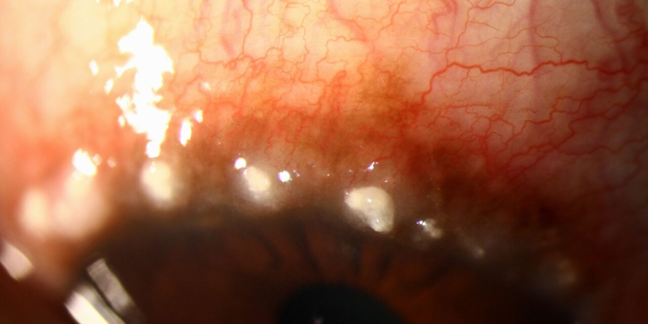
Limbal vernal keratoconjunctivitis is one of the recognized clinical patterns of vernal disease, distinct from the more commonly pictured tarsal (palpebral) form with giant cobblestone papillae, and knowing to look specifically at the limbus, not just flip the eyelid, matters because this pattern can be missed entirely if examination focuses only on the upper tarsal conjunctiva.
Two Patterns, One Disease
Vernal keratoconjunctivitis is a severe, chronic allergic eye disease that can present predominantly in one of two patterns, or as a mixed form combining features of both: the tarsal form, with giant papillae on the upper eyelid’s tarsal conjunctiva, and the limbal form, in which the pathology is centered at the corneoscleral limbus rather than the eyelid.
Recognizing which pattern, or combination, is present in a given patient matters for directing examination attention appropriately and for anticipating the specific complications each pattern is more likely to produce.
Clinical Findings in Limbal Disease
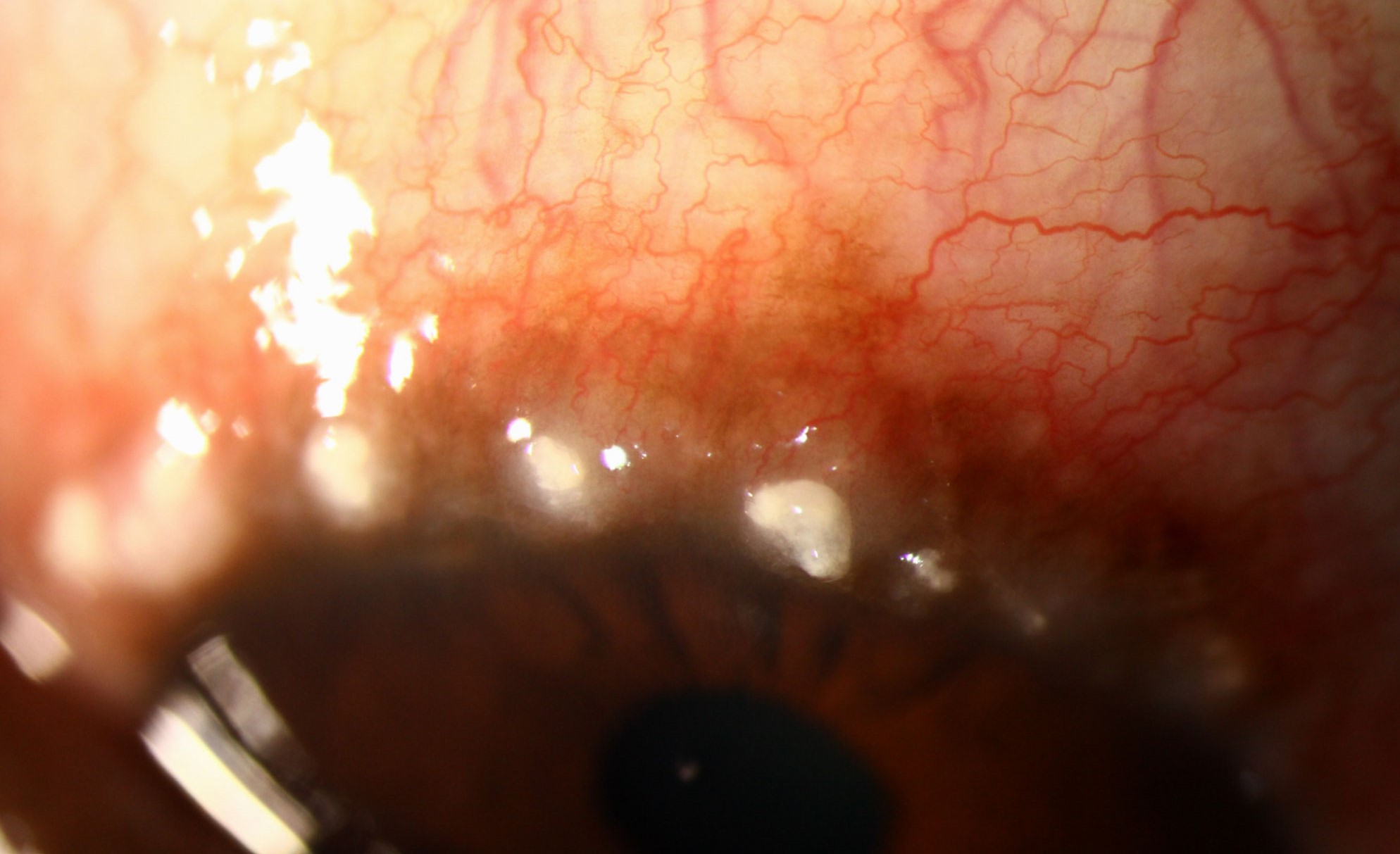
- Gelatinous, thickened, elevated limbal tissue, often most pronounced superiorly, giving the limbus a swollen, boggy appearance distinct from the normal thin limbal transition zone
- Horner-Trantas dots: small, discrete white or chalky dots scattered along the limbus, representing collections of degenerated eosinophils and epithelial cells, and considered a highly characteristic finding for active limbal vernal disease
- Limbal papillae, smaller and more numerous than the giant papillae of tarsal disease, contributing to the thickened limbal appearance
- Associated conjunctival injection and a ropy, stringy mucoid discharge, common to vernal disease more broadly regardless of the specific pattern
All-fit Slit-Lamp Adapter
Record and share exactly what you see at the slit lamp. One adapter fits any slit lamp or surgical microscope — and any smartphone.
From Choroida — the team behind this siteCorneal Complications of Limbal Disease
- Superficial punctate keratopathy, common in active disease of either pattern
- Pseudogerontoxon, a crescent-shaped, arcus-like limbal scarring that can develop after resolution of an area of limbal inflammation, sometimes persisting as a permanent marker of prior disease activity even once symptoms have settled
- Vernal (shield) ulcers can occur in limbal disease as well as tarsal disease, though the mechanism in limbal-predominant disease relates more to the toxic effect of inflammatory mediators on the adjacent cornea than to the direct mechanical trauma from giant tarsal papillae (see atopic keratoconjunctivitis shield ulcer for the related mechanism in atopic disease)
Distinguishing From Other Limbal Conditions
- The gelatinous, thickened quality and presence of Horner-Trantas dots distinguish active limbal vernal disease from a simple pinguecula or other benign limbal lesion
- A relevant history of atopy, seasonal symptom pattern (though limbal disease, like vernal disease generally, can also be perennial in some patients), and younger patient age support the diagnosis
- Associated tarsal papillae, when present concurrently, confirm the diagnosis further, though their absence does not exclude limbal-predominant vernal disease
Management
General Anti-Allergic Treatment
Management follows the same overall principles as vernal keratoconjunctivitis affecting the tarsal conjunctiva, combining topical antihistamine and mast cell stabilizer combination drops for maintenance control, topical corticosteroids for acute flares, and topical calcineurin inhibitors such as cyclosporine or tacrolimus for longer-term steroid-sparing control.
Addressing Limbal-Specific Findings
- Horner-Trantas dots and gelatinous limbal thickening generally improve with adequate control of the underlying inflammatory disease and do not require separate, targeted treatment beyond the standard anti-allergic regimen
- Corneal complications, including punctate keratopathy and any shield ulcer, are managed with the same approach used in tarsal-predominant disease: aggressive control of inflammation, lubrication, and, for a non-healing shield ulcer, debridement and bandage contact lens support
Environmental and Supportive Measures
Cool compresses, allergen avoidance where identifiable, and preservative-free lubrication support overall comfort and complement the pharmacologic anti-inflammatory regimen.
Prognosis
Limbal vernal keratoconjunctivitis, like the disease overall, tends to improve with age, often significantly lessening after puberty, though some patients continue to have milder disease activity into adulthood.
Pseudogerontoxon and other residual limbal scarring can persist as a visible marker of prior disease even after active symptoms have resolved, generally without functional consequence, while active corneal complications during the disease course require the same vigilant, prompt management appropriate to vernal keratoconjunctivitis of any pattern.


Document what you see
Two smartphone imaging tools built for everyday clinic use — one for the slit lamp, one for the fundus.
From Choroida — the team behind this siteReferences
- Bonini S, Coassin M, Aronni S, Lambiase A. Vernal keratoconjunctivitis. Eye (Lond). 2004;18:345-351.
- Leonardi A. Management of vernal keratoconjunctivitis. Ophthalmol Ther. 2013;2:73-88.
- Bonini S, Bonini S, Lambiase A, et al. Vernal keratoconjunctivitis revisited: a case series of 195 patients with long-term followup. Ophthalmology. 2000;107:1157-1163.
- Leonardi A, Busca F, Motterle L, et al. Case series of 406 vernal keratoconjunctivitis patients: a demographic and epidemiological study. Acta Ophthalmol Scand. 2006;84:406-410.
