
Williams syndrome is a genetic condition caused by a microdeletion at chromosome 7q11.23 (encompassing the elastin gene, among others), producing a recognizable combination of a distinctive “elfin” facial appearance, cardiovascular disease (classically supravalvular aortic stenosis), and a characteristic outgoing, highly sociable personality.
A striking iris finding — a stellate (star-shaped) pattern most visible in blue or light-colored irides — is frequently one of the first physical clues prompting genetic evaluation in an affected infant, sometimes noticed by an alert pediatrician or family member well before the full facial and cardiac picture has become apparent.
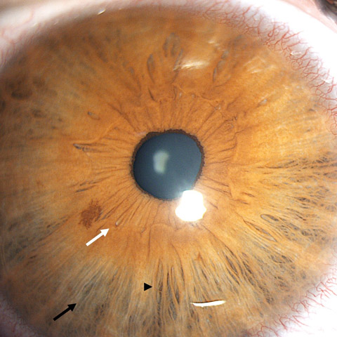
The Stellate Iris Pattern
The classic ocular finding in Williams syndrome is a stellate or lacy pattern of the iris stroma, radiating outward from the pupil in a star-like configuration, most readily appreciated in patients with blue or light-colored irides where the pattern stands out against the lighter background.
In darker irides, the same underlying stromal architecture is present but considerably harder to appreciate clinically, a genuinely important limitation of relying on this sign alone in patients with brown eyes.
This pattern reflects an underlying abnormality of iris stromal development related to the same connective tissue gene deletion (elastin) responsible for the syndrome’s vascular and connective tissue manifestations more broadly, tying together an ocular finding and the syndrome’s more medically significant systemic features through a shared molecular cause.
Because the iris finding is present from birth and does not change over time, it functions as a stable, lifelong clinical marker rather than something that needs to be tracked for progression, in contrast to some of the syndrome’s cardiovascular findings, which can genuinely worsen over childhood and require ongoing surveillance.
Other Ocular Findings
- Strabismus, particularly esotropia, occurring at a higher rate than in the general population
- Reduced stereoacuity, sometimes present even without overt strabismus
- Hyperopia (farsightedness), more common in this population than in unaffected children
- Periorbital fullness and a characteristic pattern of periorbital soft tissue contributing to the overall distinctive facial gestalt, alongside a flattened nasal bridge and full cheeks
- Blue sclera has been reported in some patients, related to the same underlying connective tissue gene involvement, though it is a less consistent and less specific finding than the stellate iris pattern
All-fit Slit-Lamp Adapter
Record and share exactly what you see at the slit lamp. One adapter fits any slit lamp or surgical microscope — and any smartphone.
From Choroida — the team behind this siteSystemic Findings
- Supravalvular aortic stenosis and other cardiovascular abnormalities — the systemic finding of greatest immediate medical significance, requiring cardiology evaluation and ongoing monitoring given the risk of progressive vascular disease
- Distinctive “elfin” facial features — full cheeks, a broad forehead, a short nose with a bulbous tip, and a wide mouth with full lips
- Intellectual disability, typically mild to moderate, combined with a characteristic, highly sociable, empathetic, and verbally engaging personality — a behavioral phenotype distinctive enough that it is often described specifically as part of the syndrome’s recognized clinical picture
- Hypercalcemia in infancy, in some affected children, requiring specific dietary and monitoring considerations during that period
- Growth abnormalities, including short stature
Differential Diagnosis
- Down syndrome — also associated with a distinctive iris finding (Brushfield spots, discussed in its own dedicated article on this site), but the two iris patterns are visually distinct (speckled ring versus radiating stellate pattern), and the two syndromes have entirely different facial gestalts, cardiac findings, and genetic causes
- Noonan syndrome — a distinct genetic condition with its own characteristic facial and cardiac features, generally without the specific stellate iris pattern of Williams syndrome
- Simple, non-syndromic stellate iris pattern — the finding can occasionally be seen as an isolated variant in individuals without Williams syndrome, though its presence alongside the syndrome’s characteristic facial, cardiac, and behavioral features is what supports the syndromic diagnosis
Diagnostic Evaluation
Genetic testing (fluorescence in situ hybridization or chromosomal microarray targeting the 7q11.23 region) confirms the diagnosis once clinically suspected based on the combination of facial, cardiac, and behavioral features, with the iris finding serving as a useful supportive clue rather than a standalone diagnostic criterion.
Given the significant cardiovascular risk, echocardiography and cardiology referral are essential parts of the initial and ongoing evaluation, alongside monitoring for hypercalcemia in infancy and developmental assessment, with cardiovascular surveillance in particular continuing at intervals throughout life rather than being a one-time evaluation at diagnosis.
Because the friendly, highly verbal, and socially engaged personality typical of Williams syndrome can sometimes mask the degree of underlying cognitive impairment on casual interaction, formal developmental and cognitive assessment is an important part of the evaluation, ensuring that educational and support planning matches the child’s actual needs rather than an impression formed from their sociable demeanor alone — a mismatch that can otherwise leave a child without services they genuinely require.
Management
There is no treatment for the underlying genetic deletion.
Ophthalmic management addresses the specific findings present in each patient — refractive correction for hyperopia, and standard strabismus management (following the same general principles discussed elsewhere on this site for esotropia and other forms of childhood strabismus) when present, with the goal of optimizing visual development and binocular function to the extent the child’s overall developmental profile allows.
Coordinated multidisciplinary care — cardiology for the cardiovascular findings, developmental and behavioral specialists, and ongoing monitoring for growth and endocrine issues — is central to comprehensive management of the syndrome as a whole, with the ophthalmic findings representing one component of a much broader care picture that typically continues throughout childhood and into adulthood.


Document what you see
Two smartphone imaging tools built for everyday clinic use — one for the slit lamp, one for the fundus.
From Choroida — the team behind this siteReferences
- Holmström G, Almond G, Temple K, et al. The iris in Williams syndrome. Archives of Disease in Childhood.
- Pober BR. Williams-Beuren syndrome. New England Journal of Medicine.
- American Academy of Ophthalmology. Basic and Clinical Science Course, Section 6: Pediatric Ophthalmology and Strabismus.
Test yourself
A few questions straight from this article.
-
Which genetic abnormality causes Williams syndrome?
Williams syndrome is caused by a microdeletion at 7q11.23, a region that encompasses the elastin gene among others, and genetic testing targets this region to confirm the diagnosis. -
What is the classic iris finding in Williams syndrome?
The classic finding is a stellate or lacy pattern of the iris stroma radiating outward from the pupil in a star-like configuration. -
In which patients is the stellate iris pattern of Williams syndrome most readily appreciated?
The pattern stands out against a lighter background, so it is hardest to appreciate in darker irides even though the same stromal architecture is present, a real limitation of relying on the sign alone. -
What links the stellate iris pattern to the vascular disease of Williams syndrome?
The iris stromal abnormality reflects the same connective tissue gene deletion, elastin, that underlies the syndrome's vascular and connective tissue manifestations more broadly. -
How does the stellate iris pattern behave over a patient's lifetime?
The iris finding is present from birth and stable, so it serves as a lifelong marker rather than something to track, unlike the cardiovascular findings that can worsen through childhood. -
Which cardiovascular lesion is classically associated with Williams syndrome?
Supravalvular aortic stenosis is the classic cardiovascular abnormality and carries the greatest immediate medical significance, requiring cardiology evaluation and ongoing monitoring. -
Which refractive error is more common in children with Williams syndrome?
Hyperopia occurs more commonly in children with Williams syndrome than in unaffected children, alongside a raised rate of strabismus, particularly esotropia. -
How does the iris finding of Down syndrome differ from that of Williams syndrome?
Both syndromes have distinctive iris findings, but Brushfield spots appear as a speckled ring while Williams syndrome produces a radiating stellate pattern, and the syndromes differ entirely otherwise. -
How should cardiovascular assessment be scheduled in a child with Williams syndrome?
Echocardiography and cardiology referral are essential at diagnosis, and cardiovascular surveillance continues at intervals throughout life rather than being a one-time evaluation. -
Why is formal cognitive assessment important in Williams syndrome?
The friendly, highly verbal manner typical of the syndrome can hide the degree of cognitive impairment on casual interaction, so formal assessment ensures educational support matches actual needs.
