
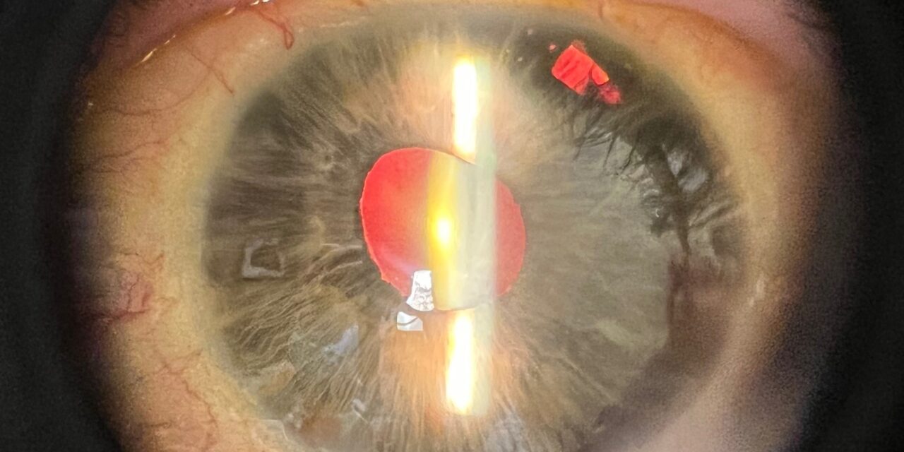
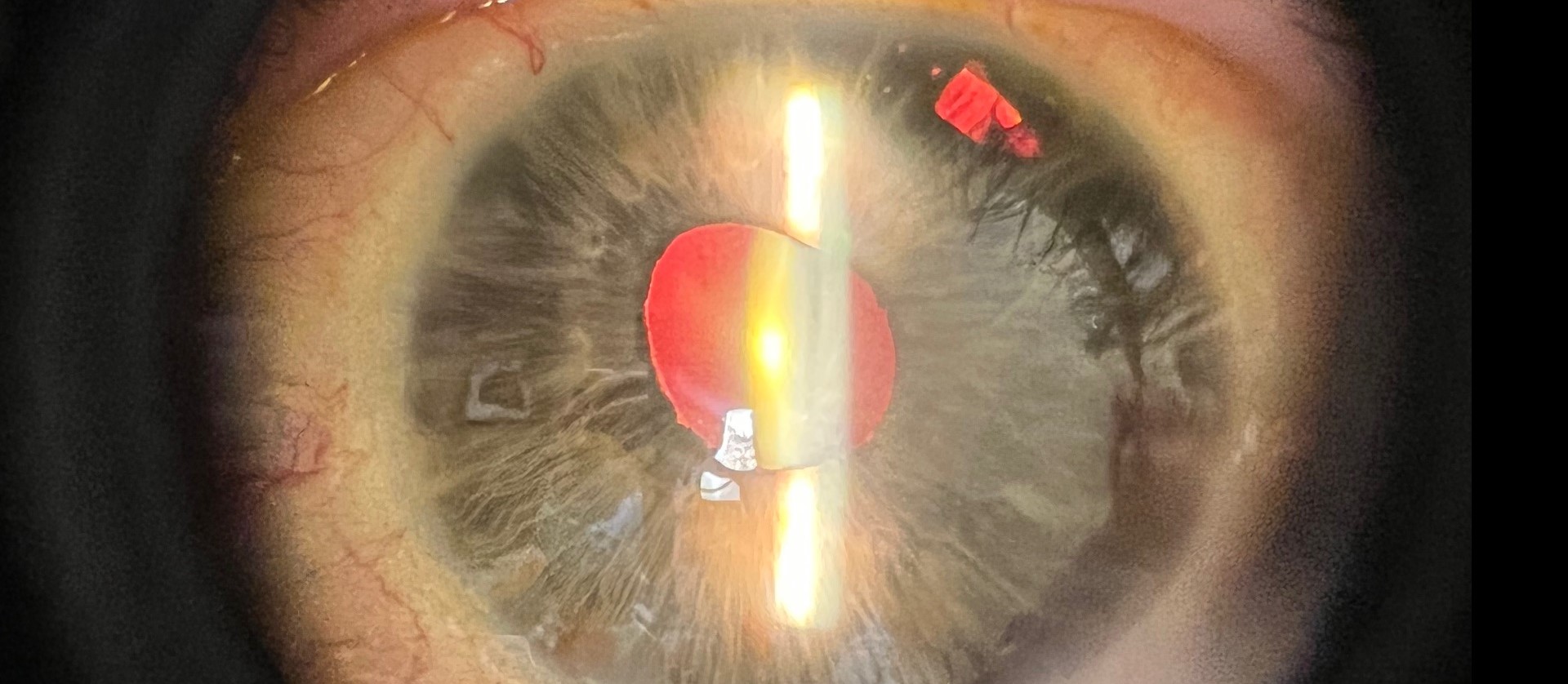
Essential iris atrophy is the most dramatically progressive member of the iridocorneal endothelial (ICE) syndrome spectrum, and watching it unfold at the slit lamp over serial visits, with the pupil slowly pulled out of shape and the iris stroma visibly thinning, gives a uniquely direct view of a disease process actively reshaping the eye in real time.
The ICE Syndrome Spectrum
Essential iris atrophy is one of three recognized clinical presentations within the iridocorneal endothelial syndrome spectrum, alongside Chandler syndrome and Cogan-Reese syndrome, all of which share a common underlying mechanism: an abnormal population of corneal endothelial cells that behaves in a way more typical of epithelial cells, proliferating and migrating across the anterior chamber angle and iris surface.
Essential iris atrophy represents the presentation with the most severe and progressive iris involvement among the three, with iris changes dominating the clinical picture more than in Chandler syndrome, where corneal edema is often more prominent, or Cogan-Reese syndrome, characterized by pigmented iris nodules.
Clinical Features
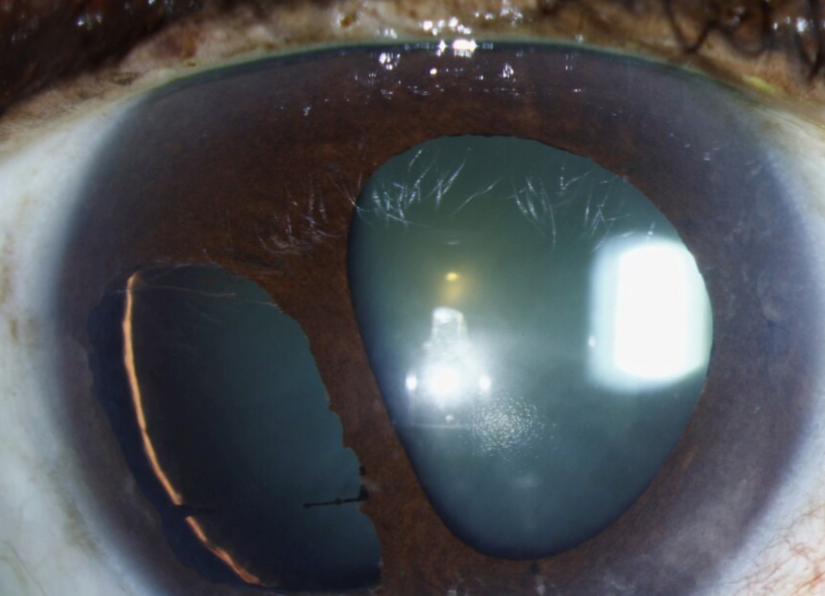
- Progressive corectopia: the pupil is gradually pulled out of its normal round shape and displaced toward the area of greatest peripheral anterior synechiae formation
- Iris stromal thinning and, in advanced cases, frank iris hole formation, sometimes multiple, as the stroma atrophies in specific areas
- Peripheral anterior synechiae, often extensive, formed by the abnormal endothelial membrane extending across the angle and contracting over time
- Secondary angle closure glaucoma, a common and significant complication, resulting from progressive angle closure by the synechiae and abnormal membrane
- Almost always unilateral, a notable and distinguishing feature from many other iris and corneal conditions, and the reason for this striking unilaterality is not fully understood
- Corneal edema can occur but is typically less prominent than in Chandler syndrome, since the corneal endothelial dysfunction here tends to be more focal
All-fit Slit-Lamp Adapter
Record and share exactly what you see at the slit lamp. One adapter fits any slit lamp or surgical microscope — and any smartphone.
From Choroida — the team behind this siteWhy the Course Is So Distinctively Progressive
The abnormal endothelial cell membrane in essential iris atrophy actively contracts over time as it spreads across the angle and iris surface, and this ongoing contractile process is what produces the continued, visible progression of corectopia and iris thinning seen on serial examination, distinguishing this from most other iris conditions, which tend to be either static or only very slowly changing.
Diagnosis
- Slit-lamp examination documenting the specific pattern and degree of corectopia, iris thinning, and any iris hole formation
- Specular or confocal microscopy demonstrating the characteristic abnormal endothelial cell appearance, sometimes described as having a “dark epithelial-like” pattern distinct from normal endothelium
- Gonioscopy to assess the extent of peripheral anterior synechiae and angle involvement
- Intraocular pressure monitoring, given the strong association with secondary glaucoma
- Serial photographic documentation is valuable for tracking the progressive nature of the condition over time
Management
Managing Secondary Glaucoma
Glaucoma management is central to the care of essential iris atrophy, since this complication is common and can be the dominant threat to vision.
- Medical therapy with aqueous suppressants is used first-line
- Surgical intervention, including glaucoma drainage device implantation, is often eventually needed given the typically progressive nature of the angle closure, with trabeculectomy generally less favored given the abnormal angle and iris anatomy
Managing Corneal Involvement
When corneal edema develops, endothelial keratoplasty can be considered, though outcomes can be complicated by the ongoing abnormal endothelial cell activity characteristic of this condition, which may continue to affect graft tissue over time.
Monitoring Progression
Given the characteristically progressive nature of essential iris atrophy, ongoing surveillance for further corectopia, angle closure, and pressure elevation is important even after initial diagnosis and treatment, since the underlying process does not necessarily stabilize on its own.
Prognosis
Essential iris atrophy tends to have a more challenging long-term course than the other ICE syndrome variants, given the combination of progressive structural iris change and a strong tendency toward secondary glaucoma that can be difficult to control.
With vigilant, sustained glaucoma management and appropriate treatment of any corneal decompensation as it arises, many patients maintain useful vision in the affected eye, though ongoing, active monitoring rather than a one-time evaluation is essential given the progressive nature of this specific condition.


Document what you see
Two smartphone imaging tools built for everyday clinic use — one for the slit lamp, one for the fundus.
From Choroida — the team behind this siteReferences
- Shields MB. Progressive essential iris atrophy, Chandler’s syndrome, and the iris nevus (Cogan-Reese) syndrome: a spectrum of disease. Surv Ophthalmol. 1979;24:3-20.
- Alvarado JA, Underwood JL, Green WR, et al. Detection of endothelial cell membrane antigens in the iridocorneal endothelial (ICE) syndrome. Trans Am Ophthalmol Soc. 1992;90:83-94.
- Laganowski HC, Kerr Muir MG, Hitchings RA. Glaucoma and the iridocorneal endothelial syndrome. Arch Ophthalmol. 1992;110:346-350.
- Sacchetti M, Mantelli F, Rehman A, Bonini S. Successful management of glaucoma associated with iridocorneal endothelial syndrome. BMJ Case Rep. 2013;2013:bcr2012007844.
