
Iris nevus versus melanoma is a distinction every ophthalmologist has to make routinely, since pigmented iris lesions are common findings on slit-lamp examination, and while the great majority are entirely benign nevi, recognizing the specific features that raise concern for malignant transformation is essential for identifying the minority that need closer surveillance or intervention.
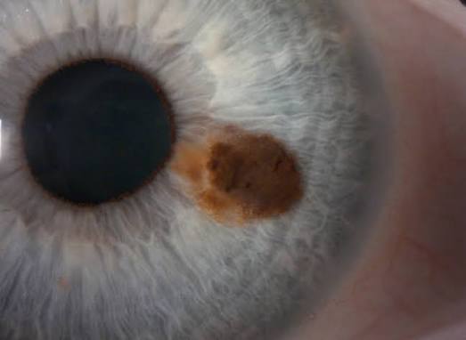
Why Most Pigmented Iris Lesions Are Benign
Iris nevi are common, benign melanocytic lesions found in a substantial proportion of the general population, and the vast majority remain entirely stable throughout a patient’s lifetime, never growing or causing any complication, which is why the default management for a typical, low-risk iris pigmented lesion is observation rather than intervention.
Iris melanoma, by contrast, is rare, but distinguishing the small subset of lesions that represent this diagnosis, or a nevus with meaningful malignant potential, from the much larger population of entirely benign nevi is the central task of iris lesion evaluation.
Risk Features Suggesting Malignancy
Several clinical features, when present, raise concern for melanoma over a benign nevus, and their presence in combination increases risk more than any single feature alone.
- Thickness: a lesion with significant elevation or thickness on slit-lamp or ultrasound biomicroscopic assessment is more concerning than a flat, uniformly pigmented lesion
- Documented growth: any objectively documented increase in size over serial examinations is one of the most significant risk indicators, which is why baseline photographic documentation of any pigmented iris lesion at first detection is so valuable
- Feathery, irregular margins: in contrast to the sharply demarcated, smooth borders typical of a stable nevus
- Prominent intrinsic vascularity: visible blood vessels within the lesion itself are an atypical and concerning finding
- Ectropion uveae: posterior pigment epithelium pulled forward onto the anterior iris surface adjacent to the lesion, suggesting an infiltrative growth pattern
- Secondary glaucoma: elevated intraocular pressure related to the lesion itself, whether from angle involvement, pigment dispersion, or mechanical effects, is a concerning associated finding
- Corectopia: distortion of the pupil shape caused by the lesion, suggesting a more infiltrative or mass-effect process
All-fit Slit-Lamp Adapter
Record and share exactly what you see at the slit lamp. One adapter fits any slit lamp or surgical microscope — and any smartphone.
From Choroida — the team behind this siteClinical Evaluation
- Careful slit-lamp examination with detailed documentation of the lesion’s size, thickness, location, color, and border characteristics
- High-quality slit-lamp photography at the initial visit, providing an objective baseline against which future growth can be reliably assessed, since subtle growth can be very difficult to judge from memory or written description alone
- Gonioscopy to assess for any angle involvement or extension
- Ultrasound biomicroscopy, particularly useful for assessing lesion thickness and any posterior extension not visible on direct examination alone
- Intraocular pressure measurement, given the association between some iris melanomas and secondary glaucoma
Monitoring Strategy
For lesions without high-risk features, periodic follow-up examination, typically at six- to twelve-month intervals initially, with repeat photographic documentation to assess for interval growth, is the standard approach, allowing genuinely benign nevi to be followed safely without unnecessary intervention.
The interval and intensity of monitoring is adjusted based on the presence of any borderline or intermediate-risk features, with closer follow-up for lesions that are not clearly low-risk but do not yet meet criteria for active concern.
When to Consider Biopsy or Treatment
- Documented growth on serial examination is generally the single most important trigger for considering active intervention
- The combination of multiple risk features, even without documented growth, may prompt earlier biopsy or treatment consideration in some cases
- Fine-needle aspiration biopsy can be considered in select cases with diagnostic uncertainty, though many decisions are made based on the clinical growth pattern and risk feature assessment without requiring tissue diagnosis
- Treatment options for confirmed or highly suspected iris melanoma include local resection (iridectomy or iridocyclectomy) for localized lesions and plaque brachytherapy, chosen based on lesion size, location, and extent
Prognosis
Iris melanoma generally has a more favorable prognosis than melanoma arising in the ciliary body or choroid, related in part to its typically smaller size and earlier detection given its visibility on routine external and slit-lamp examination.
The great majority of pigmented iris lesions encountered in practice remain benign nevi throughout follow-up, and the systematic risk-feature-based approach to evaluation and monitoring allows the rare true melanoma to be identified and treated while sparing the many patients with entirely benign lesions from unnecessary anxiety or intervention.


Document what you see
Two smartphone imaging tools built for everyday clinic use — one for the slit lamp, one for the fundus.
From Choroida — the team behind this siteReferences
- Shields CL, Kaliki S, Hutchinson A, et al. Iris nevus growth into melanoma: analysis of 1611 consecutive eyes. Ophthalmology. 2013;120:766-772.
- Khan S, Finger PT, Yu GP, et al. Clinical and pathologic characteristics of biopsy-proven iris melanoma: a multicenter international study. Arch Ophthalmol. 2012;130:57-64.
- Shields JA, Shields CL. Management of posterior uveal melanoma: past, present, and future. Retina. 2012;32:1224-1233.
- Conway RM, Chua WC, Qureshi C, Billson FA. Primary iris melanoma: diagnostic features and outcome of conservative surgical treatment. Br J Ophthalmol. 2001;85:848-854.
