
Neurotrophic keratitis is corneal disease caused by impaired corneal innervation, and one of its most dangerous features is that reduced corneal sensation removes the pain signal that would normally prompt a patient to seek care, so the disease is often more advanced than the patient’s own level of discomfort would suggest, sometimes discovered only when vision has already been threatened.
Why Corneal Nerves Matter Beyond Sensation
Corneal nerves do more than transmit pain; they release trophic factors that support epithelial cell proliferation, adhesion, and normal wound healing, and they help maintain a healthy tear film through the sensory arc that drives basal tearing and blinking.
When corneal innervation is damaged, whether from a viral infection, surgery, trauma, or a systemic neurological condition, the cornea loses both its warning system and a key support for its own maintenance, and epithelial breakdown follows even without an obvious external insult.
Causes
- Herpes simplex and herpes zoster keratitis, among the most common causes given the tropism of these viruses for sensory nerves (see herpes zoster ophthalmicus)
- Diabetes, which causes a length-dependent corneal neuropathy analogous to peripheral diabetic neuropathy elsewhere
- Trigeminal nerve damage from surgery, tumor, or trauma affecting the fifth cranial nerve
- Chronic topical medication toxicity or overuse
- Contact lens overwear
- Chemical burns and other severe ocular surface injuries
- Congenital corneal anesthesia and rare hereditary sensory neuropathies
All-fit Slit-Lamp Adapter
Record and share exactly what you see at the slit lamp. One adapter fits any slit lamp or surgical microscope — and any smartphone.
From Choroida — the team behind this siteThe Mackie Classification
Neurotrophic keratitis is staged by the Mackie classification, which describes a progression of severity useful for guiding treatment.
- Stage 1: epithelial changes including irregularity, punctate keratopathy, and reduced tear film stability, without a frank epithelial defect
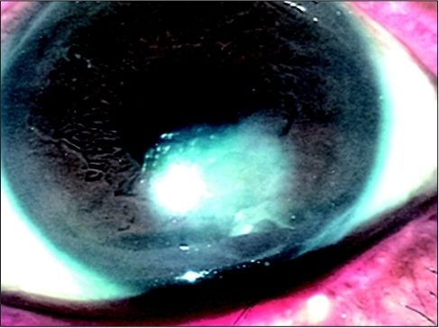
- Stage 2: a persistent epithelial defect, typically with smooth, rolled edges, often oval, and with minimal surrounding inflammation given the reduced sensory-driven inflammatory response
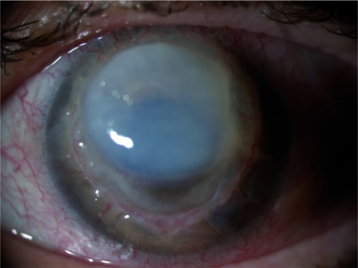
- Stage 3: stromal involvement, including stromal melting and, in severe cases, corneal perforation
Staging guides both the urgency and intensity of treatment, since stage 1 disease can often be managed conservatively while stage 3 disease requires urgent intervention to prevent perforation.
Diagnosis
- Corneal sensation testing, using a cotton wisp or, more precisely, a Cochet-Bonnet esthesiometer, to document and quantify reduced sensation, comparing the affected eye with the unaffected fellow eye when possible
- Examination for the characteristic smooth-edged, often oval, minimally inflamed epithelial defect
- A history directed at identifying the underlying cause: prior herpetic disease, diabetes, prior neurosurgical or ophthalmic surgery affecting the trigeminal nerve, or chronic medication use
Management
Supportive Measures
- Preservative-free lubrication, used frequently, to support the ocular surface in the absence of normal trophic and reflex tearing support
- Discontinuation of any toxic or unnecessary topical medications
- A bandage contact lens or moisture chamber for more significant epithelial defects
Stage-Specific Treatment
- Stage 1 disease is often managed with lubrication alone, along with treatment of any identifiable underlying cause
- Stage 2 persistent epithelial defects may need a bandage contact lens, autologous serum eye drops (which supply growth factors normally provided in part by intact corneal innervation), or, increasingly, topical recombinant human nerve growth factor
- Stage 3 disease with stromal thinning or perforation risk needs more aggressive intervention, including tissue adhesive, amniotic membrane transplantation, or tarsorrhaphy to protect the eye, and surgical repair for actual perforation
Cenegermin
Topical recombinant human nerve growth factor (cenegermin) is approved specifically for neurotrophic keratitis and has shown meaningful rates of complete corneal healing in clinical trials, representing a treatment that addresses the underlying trophic deficiency directly rather than only protecting the ocular surface while the underlying nerve damage remains unaddressed.
Surgical Options
Tarsorrhaphy, reducing the exposed corneal surface area by partially closing the eyelids, protects a non-healing or high-risk cornea, and amniotic membrane transplantation provides both mechanical protection and biological support for epithelial healing in more resistant cases.
Prognosis
Mild, early-stage disease generally responds well to supportive care and treatment of the underlying cause.
More advanced disease, particularly with stromal involvement, carries a real risk of scarring, vascularization, and perforation, and even with successful healing, the underlying reduced corneal sensation typically persists, leaving the eye at ongoing risk for recurrent breakdown, which is why long-term surveillance continues even after an acute episode resolves.


Document what you see
Two smartphone imaging tools built for everyday clinic use — one for the slit lamp, one for the fundus.
From Choroida — the team behind this siteReferences
- Mackie IA. Neuroparalytic keratitis. In: Fraunfelder FT, Roy FH, eds. Current Ocular Therapy. Philadelphia: WB Saunders; 1995.
- Bonini S, Rama P, Olzi D, Lambiase A. Neurotrophic keratitis. Eye (Lond). 2003;17:989-995.
- Dua HS, Said DG, Messmer EM, et al. Neurotrophic keratopathy. Prog Retin Eye Res. 2018;66:107-131.
- Bonini S, Lambiase A, Rama P, et al. Phase II randomized, double-masked, vehicle-controlled trial of recombinant human nerve growth factor for neurotrophic keratitis. Ophthalmology. 2018;125:1332-1343.
