
Coronavirus disease 2019 (COVID-19) is a global health crisis caused by SARS-CoV-2, an acute respiratory syndrome coronavirus.
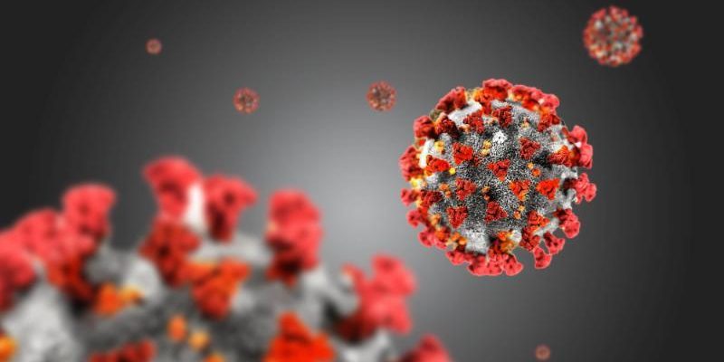
COVID-19 can cause significant ophthalmic complications, despite the majority of research and therapeutic efforts being directed towards respiratory complications.
Conjunctivitis is the most commonly reported manifestation, which may be the only symptom of SARS-CoV-2 infection in some patients.
Anterior segment manifestations of COVID-19 can affect the eyelids, ocular surface, and anterior segment, whereas posterior segment manifestations have mainly been described in case reports.
In this article, we will provide an overview of the most commonly reported retinal manifestations of COVID-19.
Microvascular Changes
Cotton wool spots and retinal microhemorrhages are the most common retinal manifestations of COVID-19. Many of these patients had preserved visual acuity and pupillary reflexes, but there have also been instances where patients developed visual field defects.
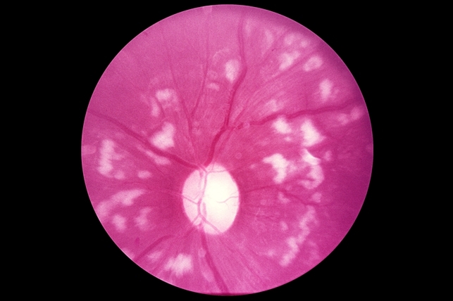
The SARS-CoV-2 infection has also been associated with new-onset paracentral acute middle maculopathy (PAMM) and acute macular neuroretinopathy (AMN), although a true relationship between these conditions and COVID-19 has yet to be established.
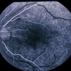
Increased tortuosity of retinal vessels is another finding that has been documented in patients with COVID-19.
However, Microvascular retinal changes are also seen in septic patients and patients with conditions like diabetic retinopathy, making it difficult to establish a causal association between SARS-CoV-2 infection and microvascular changes.
Retinal Vein Occlusion
Central retinal vein occlusion (CRVO) has been identified as an important complication of COVID-19, as early detection and treatment are necessary for improved prognosis.
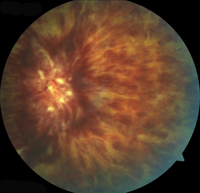
SARS-CoV-2 infection is known to cause endothelial disruption, complement activation, and inflammation, leading to a hypercoagulable state that increases the risk of thrombus formation. Decreased vision and blurred vision are the most common presenting symptoms of CRVO and can start anytime from 5 days to 6 weeks after the initial onset of fever.
Although CRVO is classically associated with risk factors like age, hypertension, glaucoma, and diabetes, COVID-19 has been shown to have a causal relationship with CRVO irrespective of patient age or comorbidities.
Because timely diagnosis and management are crucial for vision preservation, clinicians should be vigilant about monitoring for signs of CRVO in patients with a history of COVID-19.
Retinal Artery Occlusion
Central retinal artery occlusion (CRAO) is a medical emergency that can lead to complete vision loss if not treated promptly and has also been documented in the context of SARS-CoV-2 infection.
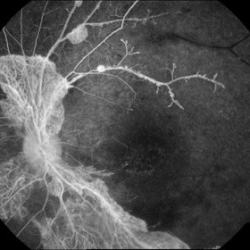
In case reports, patients developed sudden, unilateral, and painless vision loss two to six weeks after the initial onset of COVID-19 symptoms, and were found to have mild-to-significant retinal whitening on fundus exam.
However, it is important to note that most of these patients had additional underlying conditions like hypertension, obesity, and coronary artery disease, which may have placed them at a higher risk of developing CRAO.
Regardless, because rapid identification and treatment are necessary to restore visual acuity, clinicians should consider CRAO in patients with a history of COVID-19 who present with sudden and painless vision loss.


Document what you see
Two smartphone imaging tools built for everyday clinic use — one for the slit lamp, one for the fundus.
From Choroida — the team behind this siteReferences
1. Cascella M, Rajnik M, Aleem A, Dulebohn SC, Di Napoli R. Features, Evaluation, and Treatment of Coronavirus (COVID-19). In: StatPearls. StatPearls Publishing; 2022. Accessed March 12, 2022.
2. WHO Coronavirus (COVID-19) Dashboard. Accessed March 12, 2022.
3. Sen M, Honavar SG, Sharma N, Sachdev MS. COVID-19 and Eye: A Review of Ophthalmic Manifestations of COVID-19. Indian J Ophthalmol. 2021;69(3):488-509. doi:10.4103/ijo.IJO_297_21
4. Bertoli F, Veritti D, Danese C, et al. Ocular Findings in COVID-19 Patients: A Review of Direct Manifestations and Indirect Effects on the Eye. J Ophthalmol. 2020;2020:4827304. doi:10.1155/2020/4827304
Test yourself
A few questions straight from this article.
-
Which ocular manifestation is the most commonly reported in COVID-19?
Conjunctivitis is the most commonly reported ocular manifestation of COVID-19 and may be the only symptom of SARS-CoV-2 infection in some patients. -
Which retinal findings are the most commonly reported in COVID-19?
Cotton wool spots and retinal microhemorrhages are the commonest retinal manifestations, often with preserved visual acuity and pupillary reflexes. -
What is the status of the reported link between COVID-19 and paracentral acute middle maculopathy?
New-onset paracentral acute middle maculopathy and acute macular neuroretinopathy have been associated with SARS-CoV-2, but a true causal relationship is not yet established. -
Which retinal vascular change has been documented in patients with COVID-19?
Increased tortuosity of the retinal vessels is another documented finding in patients with COVID-19, alongside cotton wool spots and microhemorrhages. -
Why is it hard to attribute retinal microvascular changes directly to SARS-CoV-2 infection?
Identical microvascular retinal changes are seen in septic patients and in conditions such as diabetic retinopathy, which confounds any causal association. -
By what mechanism does SARS-CoV-2 infection predispose to retinal vein occlusion?
SARS-CoV-2 causes endothelial disruption, complement activation and inflammation, producing a hypercoagulable state that raises the risk of thrombus formation. -
Within what window after the onset of fever can COVID-related central retinal vein occlusion present?
Decreased and blurred vision are the commonest presenting symptoms of COVID-related central retinal vein occlusion, starting anywhere from 5 days to 6 weeks after fever begins. -
How does COVID-19 relate to the classical risk factors for central retinal vein occlusion?
Although vein occlusion is classically tied to age, hypertension, glaucoma and diabetes, COVID-19 has been shown to have a causal relationship regardless of age or comorbidity. -
How did COVID-associated central retinal artery occlusion present in the reported cases?
Patients developed sudden, unilateral and painless vision loss two to six weeks after COVID-19 symptoms began, with mild to significant retinal whitening on fundus examination. -
What limits the interpretation of reported COVID-associated retinal artery occlusion cases?
Most of these patients carried additional conditions such as hypertension, obesity and coronary artery disease, which may themselves have raised the risk of arterial occlusion.
