
Microphthalmia coloboma frequently occur together, since both arise from disruption of the same early developmental process, the closure of the embryonic fissure, and the combination should prompt a search for an underlying syndrome instead of being treated as an isolated ocular finding.
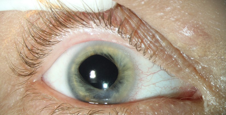
Roughly a third to half of children with these findings have an identifiable syndromic or genetic cause, and finding it changes the surveillance plan for the whole child, not just the eye.
Microphthalmia Coloboma: Developmental Basis
The optic vesicle and optic cup form early in gestation, and the embryonic fissure along the ventral surface of the developing eye normally closes by around the sixth or seventh week.
Failure of this closure leaves a coloboma, a gap that can affect the iris, ciliary body, choroid, retina, or optic nerve depending on where along the fissure closure failed, and disruption of overall globe growth around the same period produces microphthalmia, a smaller than normal eye.
The two conditions share developmental timing and often share an underlying genetic cause, which is why they are considered together as part of a spectrum along with anophthalmia, the complete absence of the eye.
Clinical Spectrum
- Isolated iris coloboma, often incidental and compatible with good vision
- Chorioretinal coloboma, which can affect vision significantly if it involves the macula or optic nerve
- Optic nerve coloboma, associated with variable but sometimes severe visual impairment
- Microphthalmia, ranging from mild to severe, sometimes with an associated orbital cyst
- Anophthalmia, the most severe end of the spectrum, requiring orbital and socket management from infancy to support normal facial bone growth
Fundus Explorer Pro
Photograph the retinal findings described here with the phone already in your pocket — 22 D optics and built-in illumination in one handheld unit.
From Choroida — the team behind this siteSyndromic Associations
A substantial number of cases are syndromic, and specific patterns of associated findings point toward particular diagnoses.
- CHARGE syndrome: coloboma, heart defects, atresia of the choanae, growth or developmental delay, genital abnormalities, and ear abnormalities, often with a CHD7 mutation
- Chromosomal abnormalities, including trisomy 13 and 18 and various deletion syndromes
- PAX6-related disorders, more classically associated with aniridia but also implicated in some microphthalmia phenotypes
- Fetal alcohol syndrome
- Congenital infections, particularly rubella and cytomegalovirus
- Isolated autosomal dominant or recessive inheritance without a broader syndrome
Workup
- A detailed family history and examination for dysmorphic features, hearing loss, cardiac disease, and developmental delay
- Genetic testing, guided by the clinical picture, ranging from targeted gene panels to chromosomal microarray or exome sequencing when the pattern is not immediately recognizable
- Cardiac evaluation, given the frequency of congenital heart disease in syndromic cases
- Hearing assessment, particularly for CHARGE syndrome and other associated conditions
- Referral to clinical genetics for children with microphthalmia or coloboma and any additional systemic finding
- Neuroimaging when optic nerve involvement or additional midline anomalies are suspected
Ocular Management
Visual potential depends on the extent and location of the coloboma and the severity of microphthalmia.
- Refractive correction and amblyopia treatment for the eye or eyes with visual potential
- Monitoring for retinal detachment, since chorioretinal coloboma carries an increased lifetime risk at the thin, abnormal margin of the defect
- Orbital and socket management in microphthalmia and anophthalmia, using conformers and expanders in infancy to stimulate normal orbital bone growth, since an underdeveloped orbit affects facial symmetry if left unaddressed
- Low-vision support and early intervention services when visual impairment is significant
Prognosis
Visual outcome varies enormously depending on which structures are involved, from normal vision with an isolated iris coloboma to severe visual impairment with extensive chorioretinal or optic nerve involvement.
The systemic prognosis depends on the underlying cause, which is why a genetics referral and syndromic workup are as important as the ophthalmic management itself for a child with microphthalmia or coloboma.


Document what you see
Two smartphone imaging tools built for everyday clinic use — one for the slit lamp, one for the fundus.
From Choroida — the team behind this siteReferences
- Chang L, Blain D, Bertuzzi S, Brooks BP. Uveal coloboma: clinical and basic science update. Curr Opin Ophthalmol. 2006;17:447-470.
- Verma AS, Fitzpatrick DR. Anophthalmia and microphthalmia. Orphanet J Rare Dis. 2007;2:47.
- Blake KD, Prasad C. CHARGE syndrome. Orphanet J Rare Dis. 2006;1:34.
- Skalicky SE, White AJ, Grigg JR, et al. Microphthalmia, anophthalmia, and coloboma and associated ocular and systemic features: understanding the spectrum. JAMA Ophthalmol. 2013;131:1517-1524.
