
Congenital nasolacrimal duct obstruction (CNLDO) is the most common cause of persistent tearing and discharge in infants, affecting a substantial minority of newborns, and it is reassuring in the sense that most cases resolve on their own within the first year of life without any procedure at all.
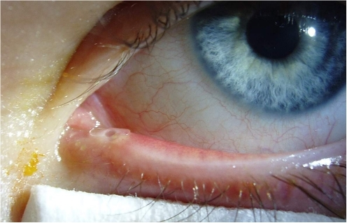
The management question is less about whether to treat and more about when watchful waiting has gone on long enough.
Why It Happens
The nasolacrimal duct develops as a solid cord of cells that normally canalizes before birth, opening at its lower end beneath the inferior turbinate at a site called the valve of Hasner.
In CNLDO, this canalization is incomplete, most often because a thin membrane persists at the valve of Hasner, leaving the duct anatomically present but functionally blocked.
Clinical Presentation
- Tearing (epiphora) that is often present from the first weeks of life
- Mucoid or mucopurulent discharge, sometimes crusting the lashes, particularly on waking
- A wet-looking eye without significant redness or photophobia, which helps distinguish it from more concerning causes of a watery, red infant eye
- Reflux of mucoid material from the punctum with pressure over the lacrimal sac
Symptoms are often bilateral, though one side may be more affected than the other.
All-fit Slit-Lamp Adapter
Record and share exactly what you see at the slit lamp. One adapter fits any slit lamp or surgical microscope — and any smartphone.
From Choroida — the team behind this siteDifferential Diagnosis
- Infantile glaucoma, which causes tearing along with photophobia, blepharospasm, corneal enlargement or haze, and needs to be excluded promptly given its urgency (see primary congenital glaucoma)
- Conjunctivitis, usually with more redness and less chronic mucoid discharge
- Punctal or canalicular agenesis, a rarer structural cause of tearing
- Congenital dacryocystocele, a bluish swelling at the medial canthus present from birth, which can become infected and needs closer attention
A calm, comfortable eye with tearing and mucoid discharge, and a normal-appearing cornea, points toward simple CNLDO rather than glaucoma.
Management
Observation and Massage
Most cases resolve spontaneously within the first year of life as the membrane at the valve of Hasner opens on its own.
Hydrostatic massage of the lacrimal sac, sometimes called Crigler massage, is taught to parents: firm downward pressure over the lacrimal sac, several times a day, aims to raise pressure within the sac and rupture the residual membrane.
Topical antibiotics are used for episodes of mucopurulent discharge but do not address the underlying obstruction.
Probing
Probing under general anesthesia is considered when symptoms persist beyond around 12 months of age, though the exact timing varies by surgeon and by how bothersome the symptoms are to the family.
Simple probing alone resolves the obstruction in a large majority of straightforward cases when performed within the first couple of years of life.
Adjunctive Procedures
When simple probing fails, or in children with a more complex or recurrent obstruction, additional procedures may be used.
- Balloon dacryoplasty, dilating the duct with a balloon catheter at the time of probing
- Nasolacrimal intubation, placing a temporary silicone tube through the system for weeks to months
- Turbinate infracture, when a large or medially positioned inferior turbinate contributes to the obstruction
Dacryocystorhinostomy
Older children or those who fail repeated probing and intubation may eventually need dacryocystorhinostomy, creating a new drainage passage directly from the lacrimal sac into the nose, bypassing the obstructed duct (see dacryocystorhinostomy).
Complications of Untreated Obstruction
Persistent obstruction predisposes to dacryocystitis, an infection of the lacrimal sac that can present with a painful, red swelling at the medial canthus and, in infants, can progress quickly and needs prompt treatment.
Chronic mucopurulent discharge can also cause secondary skin irritation and, rarely, contributes to conjunctival or corneal complications.
Prognosis
The great majority of children with CNLDO are symptom-free by their first birthday with observation and massage alone, and most of the remainder resolve with a single probing procedure.
Persistent cases usually respond to escalation through intubation or balloon dacryoplasty, and dacryocystorhinostomy is reserved for the small number who do not.


Document what you see
Two smartphone imaging tools built for everyday clinic use — one for the slit lamp, one for the fundus.
From Choroida — the team behind this siteReferences
- Paul TO, Shepherd R. Congenital nasolacrimal duct obstruction: natural history and the timing of optimal intervention. J Pediatr Ophthalmol Strabismus. 1994;31:362-367.
- Repka MX, Chandler DL, Beck RW, et al. Primary treatment of nasolacrimal duct obstruction with probing in children younger than 4 years. Ophthalmology. 2008;115:577-584.
- Kushner BJ. Congenital nasolacrimal system obstruction. Arch Ophthalmol. 1982;100:597-600.
- Pediatric Eye Disease Investigator Group. Balloon catheter dilation and nasolacrimal duct intubation for treatment of nasolacrimal duct obstruction after failed probing. Arch Ophthalmol. 2009;127:633-639.
