
Sturge-Weber syndrome is a sporadic neurocutaneous disorder in which a facial capillary malformation, a leptomeningeal vascular malformation, and ocular involvement occur together.
The eye findings are glaucoma and diffuse choroidal hemangioma, and both threaten vision.
Glaucoma can appear in infancy or years later, so patients need lifelong intraocular pressure monitoring.
Surgery carries specific risks that differ from routine glaucoma surgery, and knowing them prevents avoidable disasters.
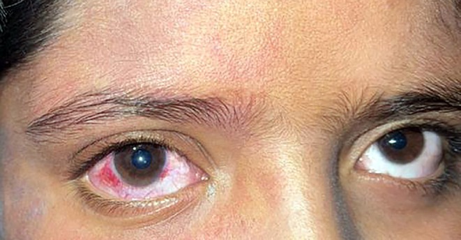
What Is Sturge-Weber Syndrome?
The condition results from a somatic mosaic activating mutation in the GNAQ gene, most often p.Arg183Gln, which affects endothelial cell signaling and leads to capillary malformations.
The syndrome comprises:
- A port-wine stain, typically in the distribution of the ophthalmic branch of the trigeminal nerve, and often involving the upper eyelid
- A leptomeningeal angioma causing seizures, hemiparesis, and developmental delay
- Ocular involvement, including glaucoma and choroidal hemangioma
Not all patients with a port-wine stain have Sturge-Weber syndrome.
The risk is higher when the stain involves the forehead or upper eyelid, or is bilateral or extensive.
Ocular Findings in Sturge-Weber Syndrome
Glaucoma
Glaucoma occurs in roughly a third to a half of patients in many series, and the estimates vary.
It is usually ipsilateral to the port-wine stain and follows a bimodal pattern.
- Early-onset glaucoma, in infancy, is related to developmental anomalies of the anterior chamber angle, and it resembles primary congenital glaucoma with buphthalmos, corneal edema, and Haab striae (see primary congenital glaucoma)
- Late-onset glaucoma, in childhood or adult life, is attributed to elevated episcleral venous pressure from arteriovenous shunting, and it behaves like open-angle glaucoma with a normal-appearing angle that shows blood in Schlemm canal
The risk of glaucoma is higher with upper eyelid involvement by the port-wine stain.
Diffuse Choroidal Hemangioma
Diffuse choroidal hemangioma appears as a thickened, red-orange choroid that gives the fundus a tomato-catsup appearance.
- It is present in a substantial proportion of patients with glaucoma
- It is often asymmetric compared with the fellow eye, and the difference in color is a clue
- It may cause hyperopia, serous retinal detachment, cystoid macular edema, and retinal degeneration
- The lesion may be subtle and may only be detected with ultrasonography, OCT, or indocyanine green angiography
Choroidal thickening and dilated choroidal vessels are seen on OCT and B-scan.
Other Ocular Features
- Dilated, tortuous conjunctival and episcleral vessels
- Iris heterochromia, with a darker iris on the affected side
- Conjunctival and episcleral hemangiomas
- Optic disc cupping from glaucoma
- Strabismus and amblyopia
Fundus Explorer Pro
Photograph the retinal findings described here with the phone already in your pocket — 22 D optics and built-in illumination in one handheld unit.
From Choroida — the team behind this siteScreening and Surveillance
Ophthalmic examination is recommended at diagnosis and at regular intervals afterward.
- Measure intraocular pressure, corneal diameter, and cup-to-disc ratio at each visit, using sedation or anesthesia in young children when necessary
- Perform gonioscopy and a dilated fundus examination
- Use OCT and B-scan to look for choroidal thickening and detachment
- Refract and treat amblyopia
- Repeat visual fields in older children and adults
Surveillance is lifelong, since late-onset glaucoma can appear in adults.
Management
Glaucoma
- Topical medications are the first step, and prostaglandin analogues, beta-blockers, and carbonic anhydrase inhibitors are used
- Angle surgery such as goniotomy or trabeculotomy may help in infantile glaucoma with angle anomalies
- Trabeculectomy and tube shunts are used for refractory disease, but they carry the risk of serious complications
The main danger is sudden ocular hypotony during surgery, which can cause choroidal effusion and suprachoroidal hemorrhage in eyes with choroidal hemangioma and high episcleral venous pressure (see choroidal detachment and suprachoroidal hemorrhage).
Precautions include controlled decompression, preoperative measures to limit effusion, prophylactic posterior sclerotomies in some centers, and careful postoperative management.
Surgery should be done by surgeons familiar with the condition.
Choroidal Hemangioma
Observation is appropriate when the eye is stable.
Subretinal fluid and macular involvement may be treated with photodynamic therapy, low-dose radiotherapy, or anti-VEGF injections in selected cases (see choroidal hemangioma).
Serous retinal detachment can appear after glaucoma surgery or with pressure fluctuation, and it may need drainage or treatment of the tumor.
Amblyopia and Visual Rehabilitation
Refractive correction and patching are used for amblyopia.
Children with hemiparesis and seizures need neurologic care, and low-vision services are provided when needed.
Skin and Neurologic Care
Pulsed dye laser treatment of the port-wine stain is provided by dermatology.
Seizure control and developmental support are managed by neurology.
Counseling and Perioperative Planning
Families should know that glaucoma can appear at any age, so pressure checks continue for life even after a normal first examination.
They should be told to report a large or cloudy cornea, tearing, photophobia, or a red and painful eye without delay.
Eye surgery and anesthesia in these patients are planned with the choroidal hemangioma in mind.
Sudden lowering of intraocular pressure, for example during paracentesis or opening of the globe, can provoke choroidal effusion, so decompression is gradual.
Patients with seizures need medication review, since some antiepileptic drugs have visual effects.
A shared care plan between ophthalmology, neurology, and dermatology avoids gaps in follow-up.
Prognosis
Vision depends on the control of glaucoma and the extent of choroidal hemangioma.
Early detection and steady pressure control preserve vision in most patients.
Refractory glaucoma and serous detachment remain the main causes of visual loss.
Care needs a team that includes ophthalmology, neurology, dermatology, and the family.


Document what you see
Two smartphone imaging tools built for everyday clinic use — one for the slit lamp, one for the fundus.
From Choroida — the team behind this siteReferences
- Shirley MD, Tang H, Gallione CJ, et al. Sturge-Weber syndrome and port-wine stains caused by somatic mutation in GNAQ. N Engl J Med. 2013;368:1971-1979.
- Sujansky E, Conradi S. Sturge-Weber syndrome: age of onset of seizures and glaucoma and the prognosis for affected children. J Child Neurol. 1995;10:49-58.
- Sullivan TJ, Clarke MP, Morin JD. The ocular manifestations of the Sturge-Weber syndrome. J Pediatr Ophthalmol Strabismus. 1992;29:349-356.
- Mantelli F, Bruscolini A, La Cava M, Abdolrahimzadeh S, Lambiase A. Ocular manifestations of Sturge-Weber syndrome: pathogenesis, diagnosis, and management. Clin Ophthalmol. 2016;10:871-878.
- Bachur CD, Comi AM. Sturge-Weber syndrome. Curr Treat Options Neurol. 2013;15:607-617.
