
Sickle cell retinopathy results from vascular occlusion by sickled red blood cells, and its proliferative form is a leading cause of vision loss in patients with sickle cell disease.
It is more common and more aggressive in hemoglobin SC disease than in homozygous SS disease, which surprises many clinicians.
Most patients keep good central vision for years, so regular screening is the best way to find eyes that need treatment.
Pathogenesis
Deoxygenated sickle hemoglobin polymerizes and deforms the red cell, which then becomes rigid and sticky.
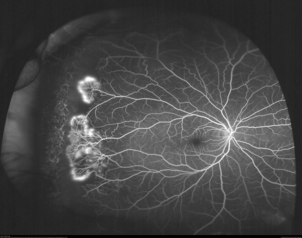
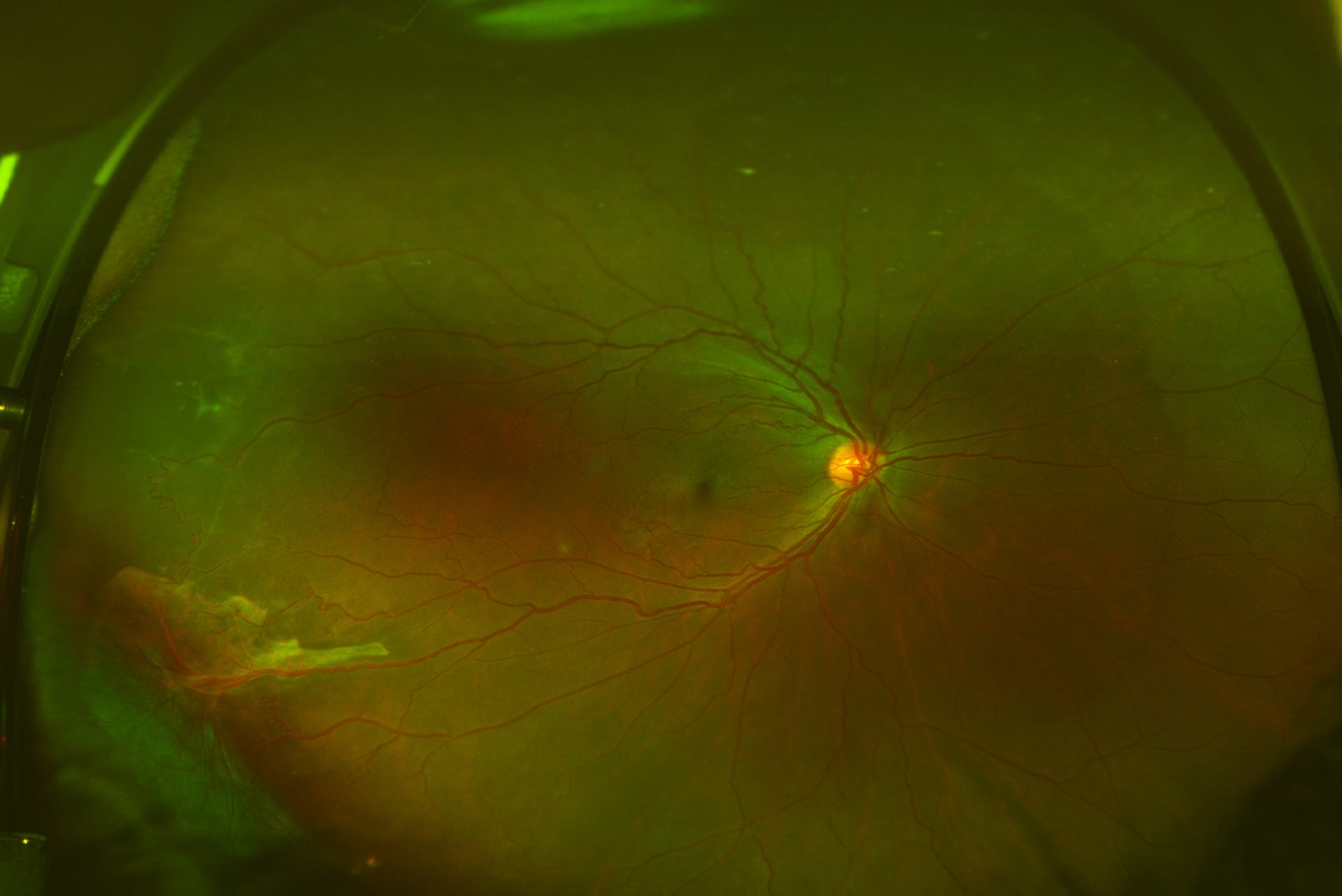
These cells occlude small peripheral retinal vessels, and the resulting ischemia is greatest at the temporal periphery, where vessels are farthest from the disc.
Ischemic retina releases VEGF, and neovascularization follows, particularly at the border of perfused and nonperfused retina.
Patients with HbSC and HbS-thalassemia have higher hematocrit and blood viscosity than SS patients, and their retinopathy is often more proliferative.
Nonproliferative Sickle Cell Retinopathy
Nonproliferative changes are common and do not require treatment.
- Salmon patch hemorrhage: a pink-orange intraretinal or preretinal hemorrhage in the midperiphery, which resolves over weeks
- Iridescent spots: refractile deposits in an old schisis cavity
- Black sunburst: a pigmented, spiculated chorioretinal scar from RPE hyperplasia after hemorrhage
- Venous tortuosity and peripheral arteriolar occlusions
- Angioid streaks in some patients (see angioid streaks)
- Macular ischemia, with enlargement of the foveal avascular zone and temporal macular thinning on OCT
Macular changes may reduce vision even without proliferation.
Fundus Explorer Pro
Photograph the retinal findings described here with the phone already in your pocket — 22 D optics and built-in illumination in one handheld unit.
From Choroida — the team behind this siteProliferative Sickle Retinopathy: The Goldberg Stages
Goldberg described a five-stage classification that is still widely used:
- Stage I: peripheral arteriolar occlusions
- Stage II: peripheral arteriovenous anastomoses at the border of perfused and nonperfused retina
- Stage III: neovascular proliferation, the classic sea fan (see sea fan neovascular frond)
- Stage IV: vitreous hemorrhage
- Stage V: tractional or combined retinal detachment
Sea fans are frond-like and may autoinfarct, becoming pale and fibrotic, which explains why some eyes regress without treatment.
In a cohort study from Jamaica, proliferative retinopathy developed most often in HbSC patients and in the second and third decades of life.
Vitreous hemorrhage in sickle cell disease is often recurrent, and traction may cause macular holes or detachment.
Macular Involvement
Central vision loss in sickle cell disease may come from macular ischemia, and it can occur without any proliferative disease.
On OCT the temporal macula thins as the inner retinal layers atrophy, and the thinning correlates with capillary dropout on OCT angiography.
Patients may notice a paracentral scotoma or reduced contrast, and they can be misdiagnosed as having an optic neuropathy.
Central retinal artery occlusion and branch occlusions occur in sickle disease, and acute vision loss in a patient with sickle disease needs urgent evaluation with hematology input.
Exchange transfusion has been used in acute occlusion, although the evidence is limited to case reports.
Screening
Dilated fundus examination is recommended at intervals, starting in childhood.
Many guidelines suggest beginning annual screening around age 10, although evidence for the best schedule is limited.
Widefield imaging and fluorescein angiography help identify peripheral ischemia and early neovascularization.
OCT and OCT angiography identify macular thinning and capillary loss, and they are helpful when acuity is discordant with the fundus appearance.
Patients with sickle trait rarely develop retinopathy, but they should be told to report any visual change, and clinicians should remember trait in unexplained retinal vascular occlusion.
Treatment
Observation
Stage I and II disease and small sea fans without hemorrhage can be observed, since spontaneous regression is common.
Laser Photocoagulation
Scatter laser to the ischemic retina or feeder vessel treatment is used for progressive sea fans and for high-risk features such as recurrent vitreous hemorrhage.
Laser in sickle retinopathy carries a risk of choroidal ischemia and bleeding, so treatment is gentle and staged.
Anti-VEGF Injections
Anti-VEGF agents can cause regression of sea fans, although evidence in sickle retinopathy comes from small series.
Vitrectomy
Vitrectomy is used for nonclearing vitreous hemorrhage and tractional or combined detachment.
Surgery requires attention to systemic status.
Perioperative and Anesthetic Precautions
Ocular surgery in patients with sickling disorders carries a higher risk of anterior segment ischemia and postoperative complications.
Precautions include:
- Coordinating with hematology, and considering exchange transfusion in selected high-risk cases
- Avoiding hypoxia, dehydration, acidosis, and hypothermia during and after surgery
- Using gentle surgical technique, with a low threshold to avoid encircling scleral buckles that compress the anterior segment circulation
- Avoiding epinephrine in the local anesthetic solution when possible
- Using short-acting gas tamponade with caution because raised IOP compromises ocular perfusion
Hyphema needs special care in patients with sickle cell disease or trait.
Sickled red cells clog the trabecular meshwork and raise IOP even at modest pressures, and optic nerve perfusion is easily lost.
Carbonic anhydrase inhibitors such as acetazolamide can promote sickling by causing systemic acidosis and should be avoided.
Systemic Treatment and the Eye
Hydroxyurea reduces pain crises and acute chest syndrome, but its effect on retinopathy is not established.
Chronic transfusion programs may lower the risk of some complications, and patients on them still need eye examinations.
Pregnancy and sickle cell disease together increase the risk of vaso-occlusive events, and ocular symptoms during pregnancy should prompt early review.
Patients should be advised to seek care for sudden vision loss, a curtain, or a sudden increase in floaters, since prompt treatment of hemorrhage and detachment changes the outcome.
Prognosis
Most patients with sickle retinopathy retain useful vision, and severe loss is uncommon in eyes treated in time.
Follow-up remains lifelong because new neovascularization can develop even after years of stable disease.


Document what you see
Two smartphone imaging tools built for everyday clinic use — one for the slit lamp, one for the fundus.
From Choroida — the team behind this siteReferences
- Goldberg MF. Classification and pathogenesis of proliferative sickle retinopathy. Am J Ophthalmol. 1971;71:649-665.
- Downes SM, Hambleton IR, Chuang EL, Lois N, Serjeant GR, Bird AC. Incidence and natural history of proliferative sickle cell retinopathy: observations from a cohort study. Ophthalmology. 2005;112:1869-1875.
- Emerson GG, Lutty GA. Effects of sickle cell disease on the eye: clinical features and treatment. Hematol Oncol Clin North Am. 2005;19:957-973.
- Elagouz M, Jyothi S, Gupta B, Sivaprasad S. Sickle cell disease and the eye: old and new concepts. Surv Ophthalmol. 2010;55:359-377.
- Yawn BP, Buchanan GR, Afenyi-Annan AN, et al. Management of sickle cell disease: summary of the 2014 evidence-based report by expert panel members. JAMA. 2014;312:1033-1048.
