
Enucleation and evisceration are the two surgical techniques for removing an eye, and the distinction between them, whether the outer scleral shell is preserved or removed along with the eye’s internal contents, has real implications for both surgical indication and the appearance and function of the resulting socket.
These are among the more emotionally significant procedures in all of ophthalmology, since they represent an irreversible decision with major implications for a patient’s appearance and psychological wellbeing well beyond the immediate medical indication driving the surgery.
Understanding when each technique is appropriate, and how orbital implant placement fits into both, helps clarify why this decision is approached with such deliberate, individualized care rather than a single default approach applied uniformly.
Both procedures remain relatively uncommon in routine practice, reserved for a specific, comparatively small subset of severe ocular disease and trauma, but their impact on the individual patient undergoing them is considerable.
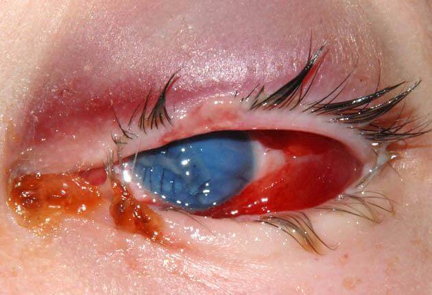
Evisceration
Evisceration removes the eye’s internal contents, including the retina, choroid, and vitreous, while preserving the outer scleral shell, extraocular muscle attachments, and the eye’s original size and shape.
Because the extraocular muscles remain attached to the preserved sclera, evisceration generally produces better postoperative motility of the resulting prosthetic eye compared to enucleation, since the muscles continue to move the intact scleral shell and, by extension, the implant and prosthesis it supports.
This technique is favored when there is no concern for intraocular malignancy, since eviscerating an eye harboring an undiagnosed tumor would fail to remove and adequately sample the tumor tissue, a genuine risk this technique specifically cannot safely accommodate.
Enucleation
Enucleation removes the entire globe, severing it from the extraocular muscles and optic nerve, and is required whenever there is known or suspected intraocular malignancy, since it allows complete removal and pathologic examination of the globe and its contents, including optic nerve margin assessment.
Because the muscles are detached from the globe during enucleation, achieving good postoperative motility requires either reattaching the muscles directly to an orbital implant or otherwise reconstructing muscle function, an additional surgical consideration not needed with evisceration.
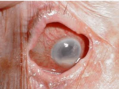
Enucleation is also generally preferred in cases of severe, unmanageable intraocular infection or in a blind, severely traumatized eye where retained ocular tissue itself poses an ongoing risk, such as with sympathetic ophthalmia, discussed in its own dedicated article on this site.
All-fit Slit-Lamp Adapter
Record and share exactly what you see at the slit lamp. One adapter fits any slit lamp or surgical microscope — and any smartphone.
From Choroida — the team behind this siteIndications
- Intraocular malignancy, including retinoblastoma or choroidal melanoma, both discussed in their own dedicated articles on this site, where enucleation is required to allow adequate pathologic staging
- A blind, painful eye, whether from end-stage glaucoma, severe uveitis, or another chronic condition, where either technique may be appropriate depending on the specific underlying diagnosis and risk of occult malignancy
- Severe ocular trauma with a non-salvageable eye, particularly when there is risk of sympathetic ophthalmia developing in the fellow eye if the severely injured eye is retained
- Severe, uncontrolled intraocular infection unresponsive to maximal medical and surgical treatment, where removing the source of infection becomes necessary
Orbital Implants and Prosthesis
An orbital implant, placed within the socket at the time of surgery, restores volume to the orbit and, when the extraocular muscles are properly attached to it, provides the foundation for movement that will later be transferred to an external prosthesis.
Various implant materials are used, ranging from inert spherical implants to porous materials that allow fibrovascular ingrowth, potentially supporting a more secure, integrated attachment for muscle reattachment and better long-term implant stability.
A custom-fitted ocular prosthesis, created and fitted by a specialized ocularist once the socket has healed sufficiently, sits over the implant and provides the visible, cosmetic surface, and it is adjusted over time as the socket matures and changes.
Psychological and Practical Considerations
Preoperative counseling addressing the emotional impact of losing an eye, and realistic expectations about prosthetic appearance and eye movement, is an essential part of preparing a patient for either procedure, not an optional addendum to the purely surgical discussion.
Support resources, including connection with others who have undergone the same procedure, can be genuinely valuable for patients adjusting to this significant, permanent change, and referral to appropriate psychological support should be offered when needed.
Coordination with an experienced ocularist from early in the process, rather than only after the socket has fully healed, also helps set realistic expectations about the timeline and appearance of the final prosthetic result.


Document what you see
Two smartphone imaging tools built for everyday clinic use — one for the slit lamp, one for the fundus.
From Choroida — the team behind this siteReferences
- American Academy of Ophthalmology. Basic and Clinical Science Course, Section 7: Oculofacial Plastic and Orbital Surgery.
- Timothy NH, Freilich DE, Linberg JV. Evisceration versus enucleation from the anaplastologist’s perspective: cosmetic outcomes. Ophthalmic Plastic and Reconstructive Surgery.
- Custer PL, Kennedy RH, Woog JJ, et al. Orbital implants in enucleation surgery: a report by the American Academy of Ophthalmology. Ophthalmology.
