
Case Study
A 17-year-old male presented with progressive bilateral drooping of the eyelids since early childhood.
His parents noted that he had difficulty keeping his eyes open, especially in dim lighting, and frequently adopted a chin-up posture to compensate.
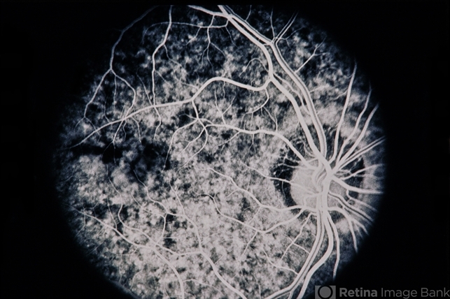
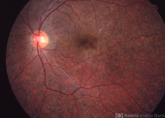
He also had a history of muscle weakness and fatigue, as well as difficulty with eye movements. Fundoscopic examination revealed pigmentary retinopathy.
A cardiac evaluation uncovered conduction abnormalities, and genetic testing confirmed a mitochondrial DNA deletion, leading to a diagnosis of Kearns-Sayre Syndrome (KSS) with associated ptosis.
The patient was managed with symptomatic treatment, including ptosis crutches and cardiac monitoring, and was counseled on the progressive nature of his condition.
Disease Entity
Kearns-Sayre Syndrome (KSS) is a rare mitochondrial disorder characterized by a triad of:
- Progressive External Ophthalmoplegia (PEO)
- Pigmentary Retinopathy
- Onset before 20 years of age
Ptosis, a hallmark feature of the disease, results from progressive dysfunction of the extraocular muscles due to mitochondrial DNA deletions.
The condition often affects multiple organ systems, making early diagnosis and monitoring crucial.
Fundus Explorer Pro
Photograph the retinal findings described here with the phone already in your pocket — 22 D optics and built-in illumination in one handheld unit.
From Choroida — the team behind this sitePathophysiology
KSS is a mitochondrial disorder caused by deletions in mitochondrial DNA (mtDNA), leading to impaired oxidative phosphorylation.
This results in energy deficits, particularly in high-energy-demand tissues such as:
- Extraocular Muscles: Leading to progressive ophthalmoplegia and ptosis.
- Retina: Causing pigmentary retinopathy.
- Cardiac Tissue: Resulting in conduction abnormalities.
- Skeletal Muscle: Causing generalized weakness.
Ptosis in KSS is primarily due to myopathic involvement of the levator palpebrae superioris rather than a neurological defect.
Epidemiology
- Age of Onset: Symptoms typically appear before 20 years of age.
- Prevalence: Rare; estimated at 1 in 100,000 individuals.
- Sex: No significant gender predilection.
Clinical Features
Ocular Findings
- Bilateral Ptosis: Usually progressive and severe.
- Progressive External Ophthalmoplegia (PEO): Limited ocular motility with sparing of pupillary function.
- Pigmentary Retinopathy: Similar to retinitis pigmentosa, presenting with a “salt-and-pepper” fundus appearance.
Systemic Findings
- Cardiac Conduction Defects: Increased risk of heart block and sudden cardiac death.
- Muscle Weakness: Proximal muscle involvement leads to fatigue.
- Endocrine Abnormalities: Including diabetes mellitus and growth hormone deficiency.
- Neurological Symptoms: Ataxia, hearing loss, and cognitive impairment in some cases.
Diagnostic Evaluation
Ophthalmic Assessment
- Slit-lamp Examination: To assess ptosis severity.
- Fundus Examination: Detects pigmentary retinopathy.
Systemic Testing
- Electrocardiogram (ECG): Essential for detecting conduction defects.
- Serum Lactate and Pyruvate: Elevated due to mitochondrial dysfunction.
- Muscle Biopsy: Shows ragged-red fibers indicative of mitochondrial disease.
- Genetic Testing: Identifies mtDNA deletions.
Differential Diagnosis
- Myasthenia Gravis: Fluctuating ptosis with positive response to acetylcholinesterase inhibitors.
- Chronic Progressive External Ophthalmoplegia (CPEO): Similar to KSS but lacks systemic involvement.
- Oculopharyngeal Muscular Dystrophy: Later onset with dysphagia.
- Congenital Ptosis: Static rather than progressive.
Management
Ocular Treatment
- Ptosis Crutches: Non-surgical option to assist eyelid elevation.
- Surgical Ptosis Repair: Considered in severe cases but may be ineffective due to weak levator function.
Systemic Treatment
- Cardiac Pacemaker: Recommended for those with conduction abnormalities.
- Coenzyme Q10 and Riboflavin: Mitochondrial support therapy with limited evidence.
- Physiotherapy: Helps maintain muscle strength.

Prognosis
- Progressive Nature: Symptoms worsen over time, but life expectancy depends largely on cardiac complications.
- Regular Monitoring: Essential for early intervention, especially for cardiac issues.
Conclusion
Kearns-Sayre Ptosis is a defining feature of Kearns-Sayre Syndrome, a mitochondrial disorder affecting multiple organ systems.
Early diagnosis and multidisciplinary management are critical to improving quality of life.


Document what you see
Two smartphone imaging tools built for everyday clinic use — one for the slit lamp, one for the fundus.
From Choroida — the team behind this siteReferences
- DiMauro S, Hirano M. Pathogenesis and treatment of Kearns-Sayre syndrome and CPEO. Neuromuscul Disord. 2006;16(5):195-202.
- Zeviani M, Muntoni F, Savarese G. Mitochondrial diseases: Molecular pathogenesis and therapeutic strategies. Brain. 2022;145(2):523-540.
- Tsao JW, Mendell JR. Mitochondrial disorders presenting with ptosis and ophthalmoplegia. Semin Neurol. 2002;22(1):33-41.
- Finsterer J. Mitochondrial disorders: Neurologic, cardiac, and ophthalmologic manifestations. Acta Neurol Scand. 2019;139(6):477-488.
Test yourself
A few questions straight from this article.
-
Which triad defines Kearns-Sayre syndrome?
Kearns-Sayre syndrome is a rare mitochondrial disorder defined by progressive external ophthalmoplegia, pigmentary retinopathy, and onset before 20 years of age. -
What genetic abnormality underlies Kearns-Sayre syndrome?
Mitochondrial DNA deletions impair oxidative phosphorylation, starving high-energy tissues such as extraocular muscle, retina, cardiac tissue and skeletal muscle. -
Why does ptosis develop in Kearns-Sayre syndrome?
Ptosis in Kearns-Sayre syndrome is myopathic, reflecting mitochondrial dysfunction of the levator palpebrae superioris rather than a neurological defect. -
What is the estimated prevalence of Kearns-Sayre syndrome?
The syndrome is rare, estimated at roughly 1 in 100,000 individuals, with symptoms appearing before 20 years and no significant sex predilection. -
Which fundus appearance is typical of the retinopathy in Kearns-Sayre syndrome?
The pigmentary retinopathy resembles retinitis pigmentosa and gives a characteristic salt-and-pepper fundus appearance on examination. -
Which systemic complication most threatens life in Kearns-Sayre syndrome?
Cardiac conduction defects carry a risk of heart block and sudden cardiac death, and life expectancy in the syndrome depends largely on cardiac complications. -
What does muscle biopsy show in Kearns-Sayre syndrome?
Muscle biopsy shows ragged-red fibres indicative of mitochondrial disease, alongside elevated serum lactate and pyruvate and mtDNA deletions on genetic testing. -
Which feature separates myasthenia gravis from Kearns-Sayre ptosis?
Myasthenia gravis produces fluctuating ptosis that responds to acetylcholinesterase inhibitors, unlike the steadily progressive myopathic ptosis of Kearns-Sayre syndrome. -
Which investigation is essential at diagnosis in Kearns-Sayre syndrome?
An ECG is essential because cardiac conduction defects may be silent yet fatal, and a pacemaker is recommended once conduction abnormalities appear. -
Which non-surgical option assists eyelid elevation in Kearns-Sayre ptosis?
Ptosis crutches assist lid elevation without surgery; surgical repair is reserved for severe cases and may fail because levator function is weak.
