
Case report
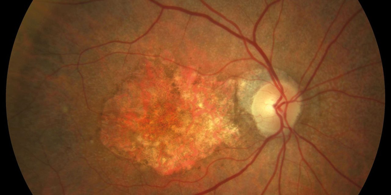
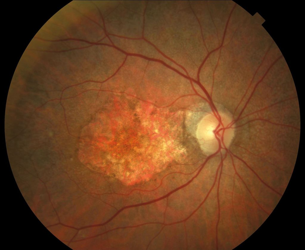
A 68-year-old female presented with a history of dry age-related macular degeneration (AMD). She presented with decreased vision in her right eye for the past two years. On examination, her visual acuity was 20/200 in the right eye and 20/20 in the left eye.

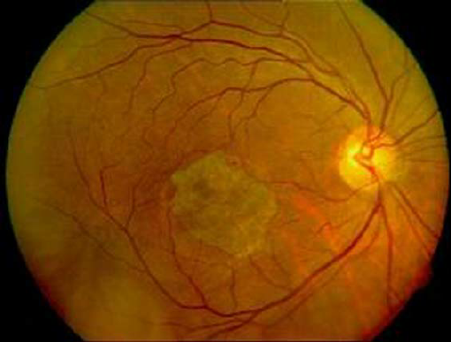
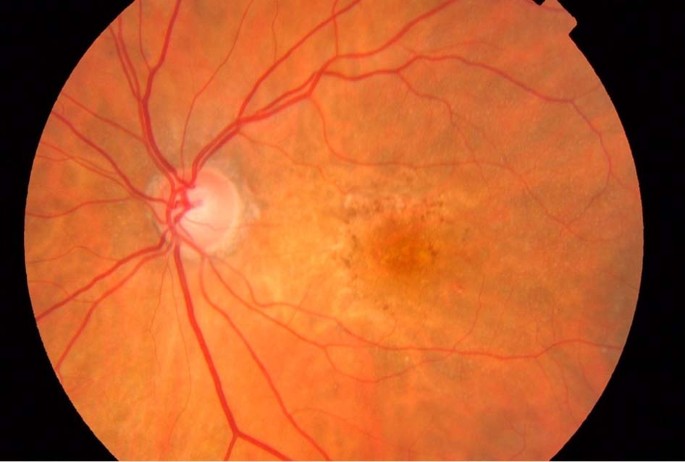
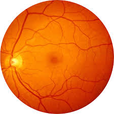
Fundus examination revealed a large area of geographic atrophy in the right eye with a well-defined border and a few drusen. Fluorescein angiography showed a large area of hypofluorescence corresponding to the GA.
Optical coherence tomography (OCT) showed a large area of retinal atrophy with a few remaining retinal layers.
Geographic atrophy DISEASE entity
Geographic atrophy is a progressive degeneration of the macula. It is an advanced stage of dry age-related macular dystrophy.
This degeneration usually starts in the perifoveal region and involves the fovea with the passage of time resulting in the central scotoma and permanent loss of vision. It is bilateral and often found in elderly people above 60 years of age.
the main complaint of a patient is gradual loss of vision, especially there is difficulty in seeing in the center of the vision inability to read books, and loss of contrast sensitivity.
Diagnosis of Geographic atrophy
Geographical atrophy can be easily diagnosed by fundoscopy however, there are some other imaging techniques that provide further details about the disease progression.
- Optical coherence tomography provides the cross-sectional mages of the retina and objectively evaluates the thickness of the retina and surrounding structures by the hypo reflectivity and hyperreflectivity pattern. This ability allows early detection of intraretinal or subretinal fluid below the RPE.
A retinal map allows the quantification of reduced retinal thickness. In the case of retinal atrophy, OCT shows a highly reflective choroidal signal due to retinal thinning and RPE hypopigmentation, allowing greater beam penetration into the choroid and greater reflectivity.
- Fundus autofluorescence is a non-invasive and standard imaging technology to visualize the retinal pigment epithelium by the hyperfluorescent and hyperfluorescent phenomenon. There is excessive lipofuscin in RPE is common in geographic atrophy.
FAF utilizes the fluorescent property of lipofuscins to create an image when exposed to a light source, the lipofuscin absorbs blue light at a specific wavelength and emits green light which is then used by a detector to record the emission signal and then creates a density map of lipofuscin.
The brighter areas represent a higher density of lipofuscin in that region which correlates well with the progression of GA. Similarly, the areas of the macula where the atrophy of RPE occurs lack fluorescent signal due to the absence of lipofuscin-containing cells.
Geographic atrophy Risk factors
- Family history & genetics
- Age: elderly people over 60 years old
- Smoking
- Hypertension & Heart disease
- Diabetes
- hypercholesterolemia
- Obesity
- Poor diet
MANAGEMENT
There is currently no treatment that can stop or reverse the progressive changes of Geographic atrophy. However, a Healthy lifestyle can lead to healthy eyes. Adapt a healthy lifestyle. Quit smoking, exercise, and eat fruits and vegetables especially dark green leafy vegetables.
Dietary supplements containing zinc, vitamins C, and E, and beta-carotene may reduce the risk of progression of AMD to its advanced stage of geographical atrophy, but these vitamins do not cure AMD.
Low vision aids can help patients to live independently by maximizing the use of the vision they have. Low-vision aids that can be used are
- Hand and stand magnifier for reading
- Telescopic lenses for distance
- Large print books and Magazines
- CCTV magnifies any printed page on a screen.
Would you have interest in taking retina images by smartphone?
Fundus photography is superior to fundus analysis as it enables intraocular pathologies to be photo-captured and encrypted information to be shared with colleagues and patients.
Recent technologies allow smartphone-based attachments and integrated lens adaptors to transform the smartphone into a portable fundus camera and Retinal imaging by smartphone.
RETINAL IMAGING BY YOUR SMARTPHONE
REFERENCES
- Fleckenstein M, Charbel Issa P, Helb HM, et al. High-resolution spectral domain-OCT imaging in geographic atrophy associated with age-related macular degeneration. Invest Ophthalmol Vis Sci. 2008;49(9):4137–4144
- Bearelly S, Chau FY, Koreishi A, Stinnett SS, Izatt JA, Toth CA. Spectral domain optical coherence tomography imaging of geographic atrophy margins. Ophthalmology. 2009;116(9):1762–1769.
- Chen JC, Fitzke FW, Pauleikhoff D, Bird AC. Functional loss in age-related Bruch’s membrane change with choroidal perfusion defect. Invest Ophthalmol Vis Sci. 1992;33(2):334–340.
- Owsley C, McGwin G, Jr., Jackson GR, Kallies K, Clark M. Cone- and rod-mediated dark adaptation impairment in age-related maculopathy. Ophthalmology. 2007;114(9):1728–1735.
- Jackson GR, Owsley C, Curcio CA. Photoreceptor degeneration and dysfunction in aging and age-related maculopathy. Ageing Res Rev. 2002;1(3):381–396.
- Scholl HPN, Bellmann C, Dandekar SS, Bird AC, Fitzke FW. Photopic and scotopic fine matrix mapping of retinal areas of increased fundus autofluorescence in patients with age-related maculopathy. Invest Ophthalmol Vis Sci. 2004;45(2):574–583.
