
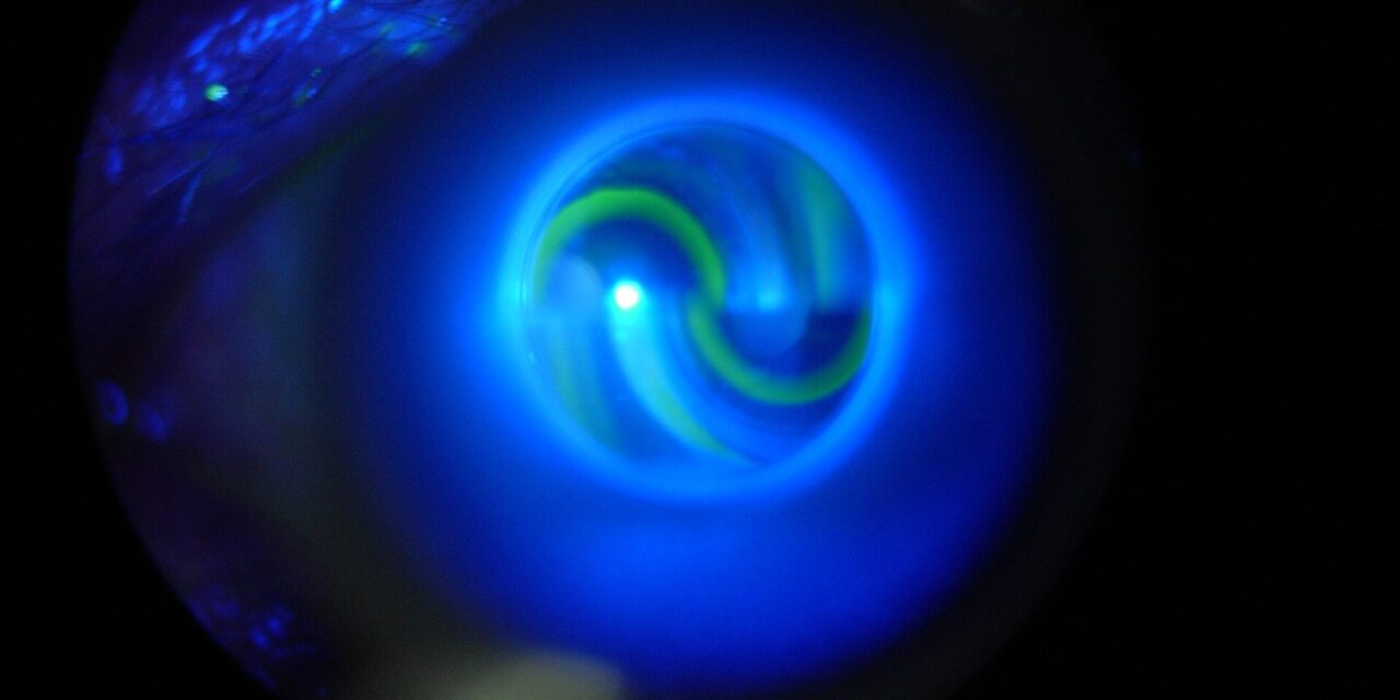
Ocular hypertension is elevated intraocular pressure occurring without the optic nerve damage or visual field loss that would define glaucoma, discussed in relation to primary open-angle glaucoma in its own dedicated article on this site, and it sits in a genuinely distinct clinical category from glaucoma itself rather than simply representing an early or mild form of the same disease.
Not every patient with ocular hypertension goes on to develop glaucoma, and understanding which specific risk factors predict a higher likelihood of progression is central to deciding which patients actually need treatment versus careful observation alone.
The Ocular Hypertension Treatment Study, the major trial that shaped modern management of this condition, transformed what had previously been a fairly uniform treat-everyone approach into the more individualized, risk-stratified framework used today.
Ocular hypertension is common enough in general eye care practice that most ophthalmologists will encounter it regularly, making a clear, individualized approach to management genuinely useful in everyday clinical decision-making rather than a rare academic consideration.
Definition
Ocular hypertension is defined as intraocular pressure above the statistically normal range, generally above 21 mmHg, in an eye with an open angle, a normal-appearing optic nerve, and a normal visual field, distinguishing it clearly from glaucoma, where structural or functional damage is already present.
This is fundamentally a statistical rather than a purely biological definition, since the normal range itself is derived from population distributions, and some eyes tolerate a pressure above this threshold indefinitely without ever developing any nerve damage.
The key clinical question in ocular hypertension is not whether the pressure is technically elevated, but whether the specific eye in front of the clinician is at meaningful risk of eventually developing glaucomatous damage from that elevated pressure.
Risk Factors for Progression to Glaucoma
- Higher baseline intraocular pressure, with risk of progression increasing roughly in proportion to how far above normal the pressure sits
- Thinner central corneal thickness, an independent risk factor identified in the major treatment trial, and one that also affects the accuracy of pressure measurement itself, discussed in relation to corneal pachymetry elsewhere on this site
- A larger cup-to-disc ratio or other subtle optic nerve features suggestive of reduced structural reserve, even when not yet meeting formal criteria for glaucomatous damage
- Older age, a family history of glaucoma, and certain other demographic and clinical factors contributing to overall risk
All-fit Slit-Lamp Adapter
Record and share exactly what you see at the slit lamp. One adapter fits any slit lamp or surgical microscope — and any smartphone.
From Choroida — the team behind this siteThe Ocular Hypertension Treatment Study
This large, influential randomized trial found that treating ocular hypertension with pressure-lowering medication reduced the risk of progression to glaucoma compared to observation alone, but also demonstrated that the great majority of untreated participants did not progress to glaucoma over the study’s follow-up period.
The trial’s risk calculator, incorporating the key risk factors identified above, allows an individualized estimate of a given patient’s five-year risk of progression, moving management away from a uniform treat-everyone approach toward one tailored to each eye’s actual calculated risk.
This individualized framework is now the standard basis for deciding which ocular hypertension patients benefit most from treatment versus careful observation, rather than treating every patient with an elevated pressure reading identically.
Management
Low-risk patients, based on a favorable combination of the factors above, are often managed with observation and regular monitoring rather than immediate treatment, avoiding the cost, side effects, and adherence burden of medication in patients unlikely to benefit meaningfully.
Higher-risk patients, particularly those with multiple risk factors or a calculated risk crossing an accepted treatment threshold, are more often started on topical pressure-lowering therapy, following similar treatment principles to established glaucoma.
Regardless of the initial management decision, regular monitoring of intraocular pressure, optic nerve appearance, and visual field remains essential, since ocular hypertension is, by definition, a condition being watched specifically for early signs that it may be evolving into glaucoma.
Patient Communication
Patients diagnosed with ocular hypertension benefit from a clear explanation that this is a risk factor and a category for monitoring, not a diagnosis of glaucoma itself, since conflating the two can cause unnecessary anxiety.
At the same time, the diagnosis should not be dismissed as entirely benign or irrelevant, since it does represent a genuine, quantifiable elevated risk that warrants ongoing attention rather than being forgotten about after the initial finding.
Revisiting the risk calculation periodically, rather than treating the initial assessment as permanent, makes sense given that risk factors such as central corneal thickness stay fixed while others, including the pressure itself and optic nerve appearance, can genuinely change over years of follow-up.


Document what you see
Two smartphone imaging tools built for everyday clinic use — one for the slit lamp, one for the fundus.
From Choroida — the team behind this siteReferences
- Kass MA, Heuer DK, Higginbotham EJ, et al. The Ocular Hypertension Treatment Study: a randomized trial determines that topical ocular hypotensive medication delays or prevents the onset of primary open-angle glaucoma. Archives of Ophthalmology.
- American Academy of Ophthalmology. Basic and Clinical Science Course, Section 10: Glaucoma.
- Gordon MO, Beiser JA, Brandt JD, et al. The Ocular Hypertension Treatment Study: baseline factors that predict the onset of primary open-angle glaucoma. Archives of Ophthalmology.
