
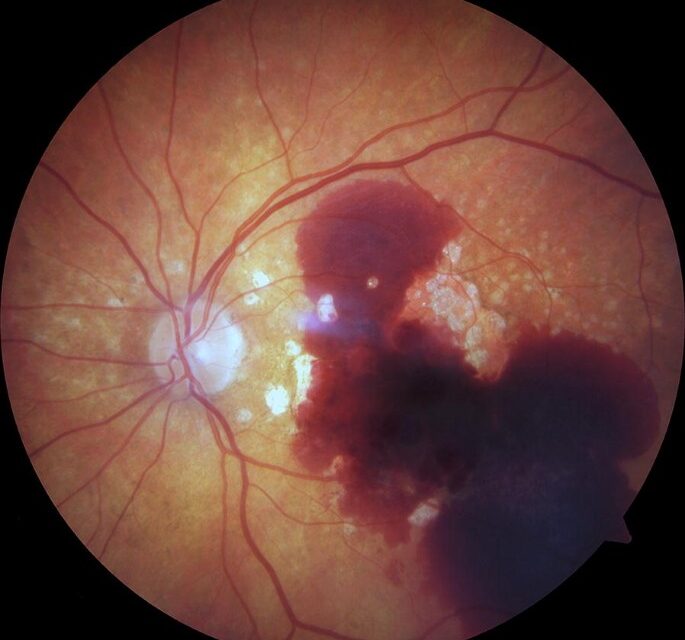
Submacular hemorrhage is bleeding trapped between the retina and the retinal pigment epithelium or beneath the RPE directly under the macula, a location that makes it disproportionately damaging compared to blood accumulating almost anywhere else in the eye.
Blood in this specific subretinal space causes injury through several distinct mechanisms simultaneously, mechanical separation, iron toxicity, and physical barrier effects, which is why even a modest volume of submacular blood can cause vision loss out of proportion to what a similar hemorrhage would cause elsewhere in the eye.
Understanding why timing matters so much in managing this condition, and why blood needs to be displaced or removed rather than simply observed and allowed to resolve on its own, is central to managing it well.
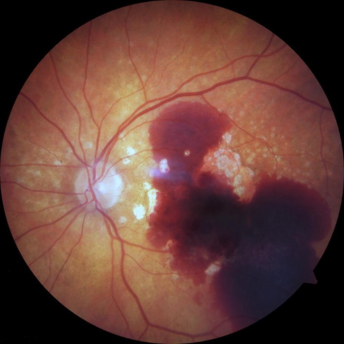
Why Location Matters So Much
The subretinal space under the macula houses the highest concentration of photoreceptors responsible for central vision, and blood in direct contact with these cells causes damage through iron and other breakdown product toxicity that begins within hours to days of the hemorrhage occurring.
Blood also physically separates the photoreceptors from the retinal pigment epithelium and underlying choroidal blood supply that normally nourishes them, compounding the direct toxic injury with a mechanical, ischemic insult to the same vulnerable tissue.
Clot contraction over the following days can add further mechanical traction and distortion to already compromised photoreceptors, meaning the damage from a submacular hemorrhage tends to worsen over time if the blood is simply left in place rather than addressed.
Causes
- Neovascular age-related macular degeneration, discussed in relation to age-related macular degeneration elsewhere on this site, one of the most common causes given how frequently fragile choroidal neovascular membranes bleed
- Retinal artery macroaneurysm, discussed in relation to macroaneurysms elsewhere on this site, which can rupture and bleed directly into the subretinal or preretinal space
- Polypoidal choroidal vasculopathy, discussed in relation to indocyanine green angiography in its own dedicated article on this site, given its characteristic polypoidal lesions’ tendency toward larger, more significant hemorrhage than typical neovascular AMD
- Trauma, including choroidal rupture, and less commonly other causes of choroidal or retinal vascular bleeding extending into the subretinal space
Fundus Explorer Pro
Photograph the retinal findings described here with the phone already in your pocket — 22 D optics and built-in illumination in one handheld unit.
From Choroida — the team behind this siteWhy Timing Matters
Photoreceptor toxicity from blood breakdown products and the mechanical effects of clot formation and contraction both worsen over time, which is the central rationale behind treating submacular hemorrhage as a time-sensitive problem rather than one that can be safely observed for an extended period before intervening.
Earlier intervention, generally within the first one to two weeks, is associated with better visual outcomes than delayed treatment in most reported series, reinforcing the general principle that displacing or removing the blood sooner limits the cumulative damage it causes.
This urgency needs to be balanced against thorough evaluation of the underlying cause, since treatment approach differs depending on that cause, but the overall message for both clinicians and patients is that this is not a condition where a wait-and-see approach serves the eye well.
Management Approaches
Intravitreal anti-VEGF injection alone is used when the hemorrhage is thin or the underlying cause is neovascular AMD without a large volume of blood requiring physical displacement, addressing the underlying neovascular drive while allowing thinner blood layers to clear on their own over time.
Pneumatic displacement, using an intravitreal gas bubble combined with patient positioning to mechanically displace blood away from the fovea, is used for thicker hemorrhages, sometimes combined with intravitreal tissue plasminogen activator to help liquefy clotted blood and facilitate its displacement.
Surgical evacuation via vitrectomy, sometimes combined with subretinal tissue plasminogen activator injected directly beneath the retina, is reserved for larger, thicker hemorrhages where pneumatic displacement alone is unlikely to be sufficient.
Prognosis
Visual outcomes depend heavily on hemorrhage size, thickness, duration before treatment, and the underlying cause, with smaller, thinner, promptly treated hemorrhages from a less aggressive underlying process generally faring better than large, longstanding hemorrhages from an aggressive neovascular process.
Even with successful blood displacement or removal, some degree of permanent photoreceptor damage from the toxic and mechanical effects already sustained is common, meaning the goal of treatment is often limiting further damage and optimizing the best achievable outcome rather than promising full visual recovery.


Document what you see
Two smartphone imaging tools built for everyday clinic use — one for the slit lamp, one for the fundus.
From Choroida — the team behind this siteReferences
- Bennett SR, Folk JC, Blodi CF, et al. Factors prognostic of visual outcome in patients with subretinal hemorrhage. American Journal of Ophthalmology.
- American Academy of Ophthalmology. Basic and Clinical Science Course, Section 12: Retina and Vitreous.
- Hillenkamp J, Surguch V, Framme C, et al. Management of submacular hemorrhage with intravitreal versus subretinal injection of recombinant tissue plasminogen activator. Graefe’s Archive for Clinical and Experimental Ophthalmology.
