
Combined rhegmatogenous-tractional retinal detachment occurs when a full-thickness retinal break coexists with active fibrovascular traction pulling on the retina, most classically in advanced proliferative diabetic retinopathy.
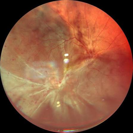
It behaves differently from either a pure rhegmatogenous detachment, discussed in relation to specific break types elsewhere on this site, or a pure tractional detachment, discussed in its own dedicated article, and it typically carries a worse prognosis and a more demanding surgical approach than either process alone.
Recognizing that both mechanisms are operating simultaneously, rather than treating the presentation as one or the other, is what actually determines how the case needs to be managed surgically.
This combined pattern is one of the more surgically challenging scenarios a vitreoretinal surgeon encounters, precisely because the two underlying problems, an anatomic break and active contractile traction, require different, sometimes competing, technical priorities during the same operation.
Mechanism
In advanced proliferative diabetic retinopathy, fibrovascular membranes growing along the posterior hyaloid and retinal surface contract over time, generating tangential and anteroposterior traction on the retina, the mechanism behind a pure tractional detachment.
When this same traction is severe enough to actually tear the retina, rather than simply distorting or elevating it without a break, subretinal fluid gains a rhegmatogenous route of entry in addition to the traction already present, and the detachment becomes a combined process.
The resulting detachment tends to progress faster than a pure tractional detachment, since rhegmatogenous fluid accumulation is generally more rapid than the slow, progressive elevation typical of traction alone.
The macula is frequently involved by the time these cases are diagnosed, since the combination of traction and an active break tends to progress before subtle early symptoms are recognized or reported.
Clinical Setting
- Advanced proliferative diabetic retinopathy with extensive fibrovascular membrane formation, the most common overall context for this presentation
- Proliferative vitreoretinopathy complicating a prior rhegmatogenous detachment repair, where contracting membranes can create new breaks or extend traction on an already compromised retina
- Penetrating trauma with both a retinal laceration and subsequent proliferative membrane formation
- Retinopathy of prematurity with advanced tractional disease, in which a superimposed break can convert a tractional detachment into a combined process
Fundus Explorer Pro
Photograph the retinal findings described here with the phone already in your pocket — 22 D optics and built-in illumination in one handheld unit.
From Choroida — the team behind this siteClinical Presentation
Patients typically present with vision loss that may be more acute or more rapidly progressive than the gradual course usually associated with a pure tractional detachment, reflecting the added rhegmatogenous component.
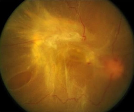
Exam findings show the characteristic taut, immobile, often star-folded or table-top configuration typical of tractional detachment, but combined with the more convex, sometimes bullous, mobile appearance characteristic of a rhegmatogenous process in the affected area.
Identifying the specific retinal break responsible for the rhegmatogenous component can be genuinely difficult amid extensive fibrovascular proliferation and hemorrhage, and it often requires careful, systematic examination, sometimes only fully clarified during surgery itself.
Surgical Approach
Pars plana vitrectomy is the mainstay of treatment, addressing both components simultaneously: removing vitreous hemorrhage, dissecting and relieving fibrovascular traction with membrane peeling or segmentation, and identifying and treating any retinal break.
Because a retinal break may not be immediately obvious amid extensive membrane and hemorrhage, careful intraoperative search, sometimes aided by perfluorocarbon liquid to help flatten and stabilize the retina during dissection, is often needed to find and treat all breaks before the case is considered complete.
Endolaser is applied around identified breaks and, often, as panretinal photocoagulation to address the underlying ischemic drive behind the fibrovascular proliferation itself, not just the mechanical consequences of that proliferation.
Long-acting gas or silicone oil tamponade is frequently used given the complexity of these cases and the meaningfully higher risk of postoperative redetachment compared to either a pure rhegmatogenous or pure tractional detachment repaired alone.
Prognosis
Combined rhegmatogenous-tractional detachments generally carry a worse anatomic and visual prognosis than either mechanism in isolation, reflecting both the severity of underlying disease typically present and the technical complexity of the repair itself.
Recurrent detachment, often from proliferative vitreoretinopathy developing after surgery, remains a significant risk, and patients need realistic counseling before surgery that more than one procedure may ultimately be needed to achieve lasting reattachment.
Optimizing systemic disease control, particularly glycemic control in diabetic patients, alongside the surgical repair, gives the best chance of a stable long-term outcome once the retina has been successfully reattached.
Close, sustained postoperative follow-up is essential given how much can still change in the months after surgery, including new membrane formation, recurrent traction, or a missed or new break declaring itself only after the initial procedure.


Document what you see
Two smartphone imaging tools built for everyday clinic use — one for the slit lamp, one for the fundus.
From Choroida — the team behind this siteReferences
- American Academy of Ophthalmology. Basic and Clinical Science Course, Section 12: Retina and Vitreous.
- Diabetic Retinopathy Vitrectomy Study Research Group. Early vitrectomy for severe proliferative diabetic retinopathy with vitreous hemorrhage. Ophthalmology.
- Charles S, Wood B. Techniques and tools for dissection of diabetic retinal membranes. Retina.
