
Before the eye is even finished developing, it briefly runs its own internal blood supply — and normally that supply disappears completely before birth.
Persistent fetal vasculature (PFV) is what happens when it doesn’t.
The fetal hyaloid vascular system nourishes the developing lens and vitreous in utero, then is supposed to regress completely by birth.
When regression fails, the leftover vascular and fibrous tissue can distort the lens, cloud the vitreous, and even drag the retina out of position.
It is one of the classic causes of leukocoria in infancy, and distinguishing it from retinoblastoma is one of the most consequential differential diagnoses in pediatric ophthalmology.
Recognizing its typical unilateral, small-eye presentation is what usually separates it from the malignancy it can otherwise resemble.
What Is Persistent Fetal Vasculature?
Persistent fetal vasculature (PFV), formerly called persistent hyperplastic primary vitreous (PHPV), is a congenital malformation from failure of the fetal hyaloid vascular system to regress normally before birth.
It is classified by the predominant location of residual tissue:
- Anterior PFV — a retrolental fibrovascular membrane, often with an associated cataract and elongated ciliary processes
- Posterior PFV — a persistent hyaloid stalk from the optic disc, sometimes with a falciform retinal fold or tractional retinal detachment
- Combined PFV — features of both anterior and posterior involvement
The vast majority of cases are unilateral and sporadic, which is one of the key clues that separates PFV from other congenital causes of leukocoria.
Epidemiology
PFV is an uncommon but well-recognized cause of congenital ocular abnormality.
- It is typically unilateral, in contrast to many other congenital ocular malformations
- Bilateral cases are less common and should prompt consideration of an associated systemic syndrome
- It is one of the classic entities in the differential diagnosis of infantile leukocoria and congenital cataract
- No strong sex predilection or major established environmental risk factor has been identified
Because PFV and retinoblastoma can both present with leukocoria in an infant, PFV’s relative frequency as a benign leukocoria cause is exactly why it cannot be assumed without a full evaluation excluding malignancy.
Fundus Explorer Pro
Photograph the retinal findings described here with the phone already in your pocket — 22 D optics and built-in illumination in one handheld unit.
From Choroida — the team behind this sitePathophysiology
The fetal hyaloid vascular system normally supports the developing lens and vitreous before regressing on a predictable timeline.
- The hyaloid artery and its associated vascular network normally regress by the third trimester, leaving the vitreous cavity clear
- In PFV, this regression is incomplete, leaving residual fibrovascular tissue in the anterior vitreous, posterior vitreous, or both
- Anterior residual tissue forms a retrolental membrane that can push against and opacify the lens, producing a cataract
- Posterior residual tissue can persist as a fibrous stalk from the optic disc, sometimes with an associated falciform fold or traction on the retina
- The affected eye is often smaller than normal (microphthalmia), reflecting the same disrupted developmental process
Because the pathology is fundamentally about residual fibrovascular tissue rather than a tumor, PFV does not carry the risk of local invasion or metastasis that makes retinoblastoma such a critical diagnosis to exclude.
Risk Factors
Established Associations
- PFV occurs sporadically in the great majority of cases, without a clear inherited pattern
- Prematurity and low birth weight have been noted in some case series, though the relationship is not as strong or consistent as with retinopathy of prematurity
Syndromic Associations
- Bilateral PFV is more likely to be associated with an underlying systemic syndrome than unilateral disease
- Rare syndromic associations have been reported, reinforcing the value of a general pediatric evaluation in bilateral cases
Because most PFV is sporadic and unilateral, an isolated finding in an otherwise healthy infant is the most common presentation clinicians will encounter.
Clinical Presentation
Symptoms and Signs Noticed by Family
- Leukocoria or an abnormal, asymmetric red reflex noticed by a parent or on newborn screening
- Strabismus, from poor vision in the affected eye
- A visibly smaller eye on the affected side in more significant cases
Examination Findings
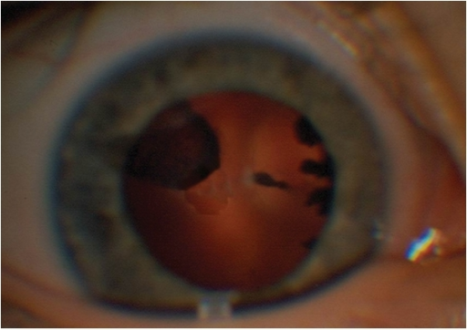
- A retrolental fibrovascular membrane visible behind the lens, often with an irregular, asymmetric appearance rather than the diffusely white reflex of a mature cataract
- Elongated ciliary processes visible once the pupil is dilated, a characteristic anterior segment finding
- A persistent hyaloid stalk extending from the optic disc toward the lens in posterior or combined disease
- Microphthalmia (a smaller globe) on the affected side, measurable and often clinically apparent
- A shallow anterior chamber, which can predispose to secondary angle-closure glaucoma over time
The irregular, often eccentric appearance of the retrolental tissue — rather than a uniform white reflex — is a useful bedside clue that favors PFV over a more centrally located retinoblastoma mass, though imaging is still required to be certain.
Diagnostic Evaluation
Examination Under Anesthesia
- Detailed anterior and posterior segment examination, typically under anesthesia in an infant, to characterize the extent of anterior and posterior involvement
- Axial length measurement to document microphthalmia
Imaging
- Ocular ultrasound to characterize the hyaloid stalk, any retinal traction or detachment, and to help exclude a calcified mass suggestive of retinoblastoma
- MRI in select cases, particularly when the diagnosis remains uncertain or posterior segment anatomy needs further clarification
Excluding Retinoblastoma
- Careful imaging assessment for calcification, which favors retinoblastoma over PFV
- A lower threshold for specialist ocular oncology referral whenever the distinction is not immediately clear
Excluding retinoblastoma is the single most important task in the workup of any infant with leukocoria — PFV is a reasonable and common explanation, but never one to assume without adequate imaging.
Differential Diagnosis
Conditions that can present similarly to PFV include:
- Retinoblastoma — the critical diagnosis to exclude, typically without microphthalmia and often with calcification on imaging
- Congenital cataract of other causes — without the characteristic retrolental membrane or elongated ciliary processes
- Coats disease — exudative retinal changes, typically presenting slightly later in childhood, usually without microphthalmia
- Retinopathy of prematurity — a clear history of prematurity with bilateral, symmetric peripheral findings
- Toxocariasis — a peripheral granuloma with a relevant exposure history
Unilaterality, microphthalmia, and the characteristic retrolental membrane appearance together make PFV recognizable, but each of these mimics still needs to be actively considered and excluded.
Management
Observation
- Appropriate for mild cases without visually significant cataract, glaucoma, or retinal traction
- Close monitoring for glaucoma, given the risk from a shallow anterior chamber
Surgical Management
- Lensectomy and removal of the retrolental membrane for visually significant anterior disease, ideally performed early to reduce amblyopia risk
- Vitrectomy for posterior disease with significant traction or hemorrhage
- Surgical timing balances the risk of amblyopia from a visually significant membrane against overall surgical risk in a very young infant
Amblyopia Management
- Aggressive amblyopia therapy (patching, optical correction) after any surgical intervention, since visual outcome depends heavily on treating the amblyopia component, not just the anatomic lesion
Even successful anatomic surgery accomplishes little without committed amblyopia therapy afterward — the two have to be planned together from the start.
Prognosis
Visual prognosis is highly variable and depends on the severity and location of disease.
- Mild anterior disease without significant cataract or traction can retain reasonably good vision, particularly with early, committed amblyopia treatment
- Posterior or combined disease with significant retinal traction or detachment carries a substantially worse visual prognosis
- Secondary glaucoma, when it develops, adds further risk to long-term visual outcome and requires its own ongoing management
Outcomes in PFV are determined as much by how early and consistently amblyopia is treated as by the success of any single surgery on the eye itself.


Document what you see
Two smartphone imaging tools built for everyday clinic use — one for the slit lamp, one for the fundus.
From Choroida — the team behind this siteReferences
- Sisk RA, Berrocal AM, Feuer WJ, Murray TG. Visual and anatomic outcomes with or without surgery in persistent fetal vasculature. Ophthalmology. 2010.
- Goel N, Kumar V, Seth A, et al. Congenital anomalies of the vitreous. Indian Journal of Ophthalmology. 2014.
- Persistent Fetal Vasculature. StatPearls, NCBI Bookshelf.
- Persistent Fetal Vasculature. EyeWiki, American Academy of Ophthalmology.
- Shastry BS. Persistent hyperplastic primary vitreous: congenital malformation of the eye. Clinical and Experimental Ophthalmology. 2009.
Test yourself
A few questions straight from this article.
-
Persistent fetal vasculature was formerly known by which name?
PFV was previously called persistent hyperplastic primary vitreous, a congenital malformation caused by failure of the fetal hyaloid vascular system to regress before birth. -
Failure of which structure to regress produces persistent fetal vasculature?
The fetal hyaloid vascular system nourishes the developing lens and vitreous in utero and should disappear completely before birth; PFV is what happens when that regression fails. -
By what point in development does the fetal hyaloid vascular system normally regress?
The hyaloid artery and its vascular network normally regress by the third trimester, leaving the vitreous cavity clear. Incomplete regression leaves residual fibrovascular tissue. -
Which anterior segment finding, visible once the pupil is dilated, is characteristic of persistent fetal vasculature?
Elongated ciliary processes drawn toward the retrolental membrane are a characteristic anterior segment sign of PFV, seen once the pupil is dilated. -
What is the typical laterality of persistent fetal vasculature?
The vast majority of PFV cases are unilateral and sporadic, and this unilaterality is a key clue separating it from other congenital causes of leukocoria. -
Which globe finding typically accompanies persistent fetal vasculature on the affected side?
The affected eye is often smaller than normal, reflecting the same disrupted developmental process; axial length measurement is used to document the microphthalmia. -
Which imaging feature favours retinoblastoma over persistent fetal vasculature in an infant with leukocoria?
Ocular ultrasound is used partly to look for calcification, which favours retinoblastoma. PFV instead shows a hyaloid stalk, retrolental membrane and a small globe. -
Which anterior segment feature of persistent fetal vasculature predisposes to secondary angle-closure glaucoma?
A shallow anterior chamber is common in PFV and can lead to secondary angle-closure glaucoma over time, so close intraocular pressure monitoring is part of even conservative management. -
What is the surgical treatment for visually significant anterior persistent fetal vasculature?
Lensectomy with removal of the retrolental membrane is performed early to limit amblyopia risk; vitrectomy is reserved for posterior disease with significant traction or hemorrhage. -
Which form of persistent fetal vasculature carries the worst visual prognosis?
Posterior or combined disease with significant traction or detachment does substantially worse, while mild anterior disease can retain reasonable vision with early, committed amblyopia therapy.
