
Not every heavy, tired-looking eyelid is ptosis — and mixing the two up leads straight to the wrong surgery.
Dermatochalasis is excess, lax eyelid skin, and it is one of the most common age-related changes seen in any eye clinic.
It is frequently confused with true ptosis, since both can make the eye look smaller or more hooded.
The distinction matters because the two conditions come from entirely different tissue problems and are corrected with different operations.
Beyond appearance, redundant skin can genuinely obstruct the superior visual field once it becomes advanced.
Recognizing dermatochalasis accurately — and separating it from ptosis, brow ptosis, and other eyelid mimics — is what leads to the right treatment plan.
What Is Dermatochalasis?
Dermatochalasis is excess, redundant skin of the upper (and sometimes lower) eyelid resulting from loss of skin elasticity, typically with age.
Its defining features include:
- Excess, often crepe-like, loose skin overhanging the eyelid margin
- A normal underlying levator muscle and normal eyelid margin position (MRD1), unlike true ptosis
- Frequent coexistence with fat prolapse (steatoblepharon), adding to the fullness of the upper lid
- A spectrum from mild cosmetic concern to significant functional obstruction of the superior visual field
Because the lid margin itself sits in a normal position, dermatochalasis is fundamentally a skin problem, not a muscle or nerve problem — that distinction is the whole basis for correctly separating it from ptosis.
Epidemiology
Dermatochalasis is extremely common and becomes progressively more prevalent with age.
- It is rare before middle age and increases steadily in prevalence from the fifth decade onward
- Both sexes are affected, though presentation and cosmetic concern patterns can differ
- A family history of early skin laxity is a recognized contributor in some patients
- It is one of the most common reasons for oculoplastic consultation in older adults
Because it is so common with normal aging, the clinical question is rarely whether some dermatochalasis is present, but whether it has become functionally or cosmetically significant enough to warrant intervention.
All-fit Slit-Lamp Adapter
Record and share exactly what you see at the slit lamp. One adapter fits any slit lamp or surgical microscope — and any smartphone.
From Choroida — the team behind this sitePathophysiology
Dermatochalasis reflects cumulative structural changes in eyelid skin and its supporting tissue.
- Chronic UV exposure and normal aging reduce dermal collagen and elastin content over time
- Loss of skin elasticity allows the thin eyelid skin to stretch and become redundant
- Weakening of the orbital septum permits orbital fat to prolapse forward, adding to upper lid fullness
- Gravitational descent of the eyebrow (brow ptosis) frequently compounds the appearance of excess upper lid skin
Because eyebrow position, orbital fat, and skin laxity all interact, a full assessment needs to account for all three rather than treating loose skin as the only variable.
Risk Factors
Established Risk Factors
- Increasing age
- Cumulative UV/sun exposure
- Smoking, associated with accelerated skin aging generally
- A family history of early or pronounced periocular skin laxity
Contributing Factors
- Chronic eye rubbing
- Thyroid eye disease or other causes of chronic eyelid edema, which can stretch skin over time
- Prior eyelid trauma or surgery affecting skin elasticity in the area
Sun protection is one of the few genuinely modifiable factors, though established dermatochalasis itself does not reverse with prevention alone.
Clinical Presentation
Symptoms
- A heavy, tired, or aged appearance of the upper eyelids, often the primary cosmetic complaint
- Superior visual field obstruction in more advanced cases, sometimes affecting activities like driving or reading
- Brow ache from chronic compensatory frontalis muscle use to lift heavy lids
- Difficulty applying makeup or wearing glasses comfortably in some patients
Examination Findings
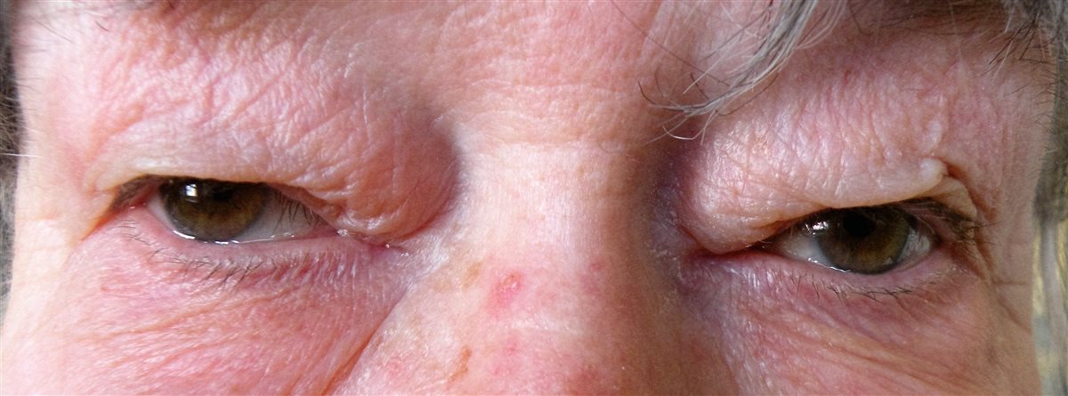
- Redundant, often crepe-textured skin overhanging the upper eyelid margin
- Normal marginal reflex distance (MRD1) and normal levator function, distinguishing it from true ptosis
- Visible fat prolapse (steatoblepharon), giving the lid a fuller, puffy appearance
- Brow position assessment to determine whether coexisting brow ptosis is contributing to the appearance
- Visual field testing with the skin taped up versus in its natural position, useful for functional/insurance documentation
A normal MRD1 with abnormal skin folding is the key exam finding that confirms dermatochalasis rather than ptosis — measure, don’t just look.
Diagnostic Evaluation
Clinical Examination
- MRD1 and levator function measurement to exclude a coexisting or mimicking ptosis
- Assessment of brow position, since brow ptosis often needs to be addressed alongside or instead of the eyelid skin itself
Functional Visual Field Documentation
- Formal visual field testing (with and without the redundant skin taped up) to document functional impairment
- External photography showing the visual axis obstruction, often required for insurance-supported functional blepharoplasty
Objective visual field documentation is what separates a functional, medically indicated blepharoplasty from a purely cosmetic one — this distinction usually determines insurance coverage.
Differential Diagnosis
Conditions that can be confused with dermatochalasis include:
- Ptosis — abnormally low MRD1 from a levator or neurologic problem, with normal-appearing skin
- Brow ptosis — the eyebrow itself has descended, pushing skin down onto the lid without primary skin redundancy
- Blepharochalasis — a rarer, recurrent inflammatory eyelid edema syndrome in younger patients, leaving thin, redundant skin after repeated attacks
- Thyroid eye disease — eyelid fullness from orbital fat and muscle expansion rather than primary skin laxity, often with proptosis or lid retraction
Distinguishing these matters because the surgical plan differs substantially — blepharoplasty for skin, brow lift for ptotic brows, and neither alone will fully correct the other’s contribution if mixed up.
Management
Observation
- Appropriate for mild dermatochalasis without functional visual impairment or significant cosmetic concern
Surgical Correction
- Upper eyelid blepharoplasty to excise redundant skin (and, when present, prolapsed fat), the definitive treatment for functionally or cosmetically significant disease
- Brow lift, performed alone or combined with blepharoplasty, when brow ptosis is a significant contributor
- Careful preoperative marking and conservative skin excision to avoid overcorrection and lagophthalmos
Non-Surgical Considerations
- Sun protection to slow further skin aging, though it does not reverse existing redundancy
- Addressing any contributing chronic eyelid edema (e.g., from thyroid eye disease) as part of overall management
Blepharoplasty remains the only truly definitive treatment — no topical or non-surgical option meaningfully reverses established skin redundancy.
Prognosis
Prognosis is excellent, and this is generally a highly satisfying condition to treat surgically.
- Upper eyelid blepharoplasty reliably improves both appearance and, when present, visual field obstruction
- Results are generally long-lasting, though the normal aging process can gradually produce some recurrence over many years
- Complications are uncommon with careful surgical technique, though overly aggressive skin removal risks lagophthalmos and ocular surface exposure
Correctly identifying whether skin, fat, brow position, or a coexisting ptosis is driving the appearance is what determines whether a single blepharoplasty fully solves the problem or leaves the patient wondering why they still look tired.


Document what you see
Two smartphone imaging tools built for everyday clinic use — one for the slit lamp, one for the fundus.
From Choroida — the team behind this siteReferences
- Dermatochalasis. StatPearls, NCBI Bookshelf.
- Damasceno RW, Cariello AJ, Cardoso EB, et al. Upper blepharoplasty with or without resection of the orbicularis oculi muscle. Ophthalmic Plastic and Reconstructive Surgery. 2011.
- Dermatochalasis. EyeWiki, American Academy of Ophthalmology.
- American Society of Plastic Surgeons. Blepharoplasty guidelines.
- Wladis EJ, Aakalu VK, Foster JA, et al. Interventions for involutional blepharoptosis: a report by the American Academy of Ophthalmology. Ophthalmology. 2018.
Test yourself
A few questions straight from this article.
-
Which tissue abnormality defines dermatochalasis?
Dermatochalasis is redundant upper (and sometimes lower) eyelid skin from age-related loss of skin elasticity, with the levator and lid margin position both normal. -
Which measurement separates dermatochalasis from true ptosis at the slit lamp?
In dermatochalasis the lid margin sits normally, so MRD1 and levator function are normal; a low MRD1 points instead to a levator or neurologic ptosis. -
What term describes the prolapsed orbital fat that often accompanies dermatochalasis?
Weakening of the orbital septum lets orbital fat prolapse forward, called steatoblepharon, adding to the puffy fullness of the upper lid. -
Which structural change allows orbital fat to prolapse forward in dermatochalasis?
The orbital septum normally holds fat back; as it weakens with age, fat comes forward and compounds the redundant skin above the lid margin. -
Which functional visual complaint arises from advanced upper lid dermatochalasis?
Redundant skin hanging over the lid margin can genuinely block the superior field, affecting tasks such as driving and reading, not just appearance. -
Why do patients with dermatochalasis often complain of brow ache?
Patients recruit the frontalis continuously to lift heavy lids out of the visual axis, and that sustained effort produces the characteristic brow ache. -
Which test documents functional impairment from dermatochalasis for insurance purposes?
Formal fields taped versus untaped, supported by external photographs of the visual axis obstruction, are what separate a functional blepharoplasty from a cosmetic one. -
Which mimic of dermatochalasis causes recurrent inflammatory eyelid edema in younger patients?
Blepharochalasis is a rarer recurrent inflammatory eyelid edema syndrome of younger patients that leaves thin, redundant skin after repeated attacks, unlike age-related dermatochalasis. -
What is the definitive treatment for functionally significant dermatochalasis?
Blepharoplasty excises the redundant skin and any prolapsed fat, and no topical or non-surgical option meaningfully reverses established skin redundancy. -
Which complication follows overly aggressive skin excision during dermatochalasis surgery?
Conservative marking and excision matter because removing too much skin leaves the lids unable to close fully, exposing the ocular surface.
