
Vitreous amyloidosis is deposition of amyloid protein within the vitreous cavity, most classically seen in hereditary transthyretin amyloidosis, and it can be the presenting feature that brings an undiagnosed systemic condition to medical attention.
Unlike most vitreous opacities, which are inert debris or blood, amyloid deposits are biologically active tissue infiltrating the vitreous structure itself, which is part of why they behave differently both clinically and on examination from other causes of vitreous haze.
An ophthalmologist recognizing the distinctive appearance of vitreous amyloid, and connecting it to the possibility of an underlying systemic amyloidosis, can meaningfully accelerate a diagnosis that might otherwise take considerably longer to reach through other specialties.
The condition is rare overall, but it is concentrated in specific populations and endemic regions where the causative TTR mutations are more prevalent, which is useful context when a patient’s ancestry or family history raises the possibility.
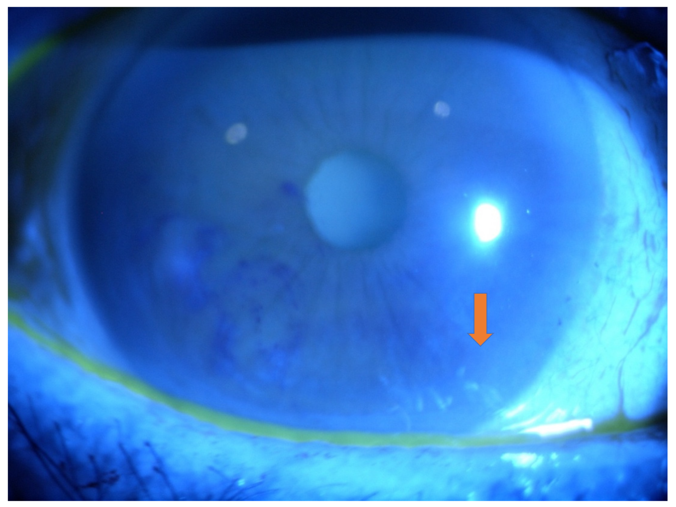
Pathophysiology
Hereditary transthyretin amyloidosis results from mutations in the TTR gene, causing the transthyretin protein to misfold and deposit as amyloid fibrils in multiple tissues throughout the body, including peripheral nerves, the heart, and the eye.
Ocular involvement occurs because transthyretin is also produced locally within the eye, by the retinal pigment epithelium and structures of the ciliary body, meaning vitreous amyloid deposition can develop somewhat independently of the systemic disease’s severity or stage.
This local production explains an important clinical point: liver transplantation, which corrects the systemic source of mutant transthyretin production in hereditary amyloidosis, does not reliably halt ongoing ocular amyloid deposition, since the eye continues producing the abnormal protein on its own.
Other ocular manifestations of the same disease process, including pupillary abnormalities, glaucoma from angle involvement, and dry eye from autonomic denervation, can accompany the vitreous findings, reflecting how broadly transthyretin amyloid can affect ocular tissue.
Clinical Presentation
- Progressive, often bilateral vision loss and floaters, developing gradually over months to years rather than appearing acutely
- A characteristic vitreous appearance often described as glass-wool or veil-like opacities, distinct from the appearance of vitreous hemorrhage or typical inflammatory debris
- Vitreous opacities that classically do not settle inferiorly the way blood or ordinary debris does, instead remaining suspended in a web-like pattern throughout the vitreous cavity
- Associated systemic findings in hereditary cases, including peripheral neuropathy, autonomic dysfunction, and cardiomyopathy, which may already be present or may not yet be diagnosed at the time of the ocular presentation
Fundus Explorer Pro
Photograph the retinal findings described here with the phone already in your pocket — 22 D optics and built-in illumination in one handheld unit.
From Choroida — the team behind this siteWhy Recognition Matters
Vitreous amyloidosis can be the first clinically apparent manifestation of hereditary transthyretin amyloidosis in some patients, particularly in later-onset or atypical presentations of the systemic disease.
A patient presenting with this characteristic vitreous appearance, especially with a family history of neuropathy, cardiomyopathy, or unexplained systemic illness, warrants referral for systemic evaluation and genetic testing rather than being managed as an isolated ophthalmic finding.
Early systemic diagnosis has real treatment implications now that disease-modifying therapies for hereditary transthyretin amyloidosis exist, making the ophthalmologist’s role in recognizing the ocular sign potentially significant for the patient’s overall prognosis, not just their vision.
Diagnostic Evaluation
The diagnosis is often suspected from the distinctive clinical appearance of the vitreous opacities on exam, particularly their veil-like quality and resistance to settling.
Vitreous biopsy, typically obtained at the time of therapeutic vitrectomy, can confirm the diagnosis histologically with amyloid-specific staining, providing definitive tissue confirmation when needed.
Genetic testing for TTR mutations and a systemic workup, coordinated with cardiology and neurology given the multisystem nature of hereditary transthyretin amyloidosis, are appropriate once the ocular finding raises suspicion.
Management
Pars plana vitrectomy is the primary treatment for visually significant vitreous amyloid, removing the opacified vitreous and typically providing meaningful visual improvement, at least temporarily.
Recurrence of vitreous amyloid deposition after vitrectomy is common, since local ocular production of the abnormal protein continues regardless of the systemic disease’s treatment status, and repeat vitrectomy is sometimes needed over the years that follow.
Management of the underlying systemic disease, including newer transthyretin-stabilizing or gene-silencing therapies, is coordinated with the patient’s systemic specialists, since these treatments primarily target systemic organ involvement rather than reliably halting ocular deposition.
Genetic counseling for the patient’s family also deserves attention given the autosomal dominant inheritance pattern typical of hereditary transthyretin amyloidosis, since at-risk relatives may benefit from their own baseline evaluation and testing.


Document what you see
Two smartphone imaging tools built for everyday clinic use — one for the slit lamp, one for the fundus.
From Choroida — the team behind this siteReferences
- Sandgren O. Ocular amyloidosis, with special reference to the hereditary forms with vitreous involvement. Survey of Ophthalmology.
- American Academy of Ophthalmology. Basic and Clinical Science Course, Section 12: Retina and Vitreous.
- Kawaji T, Ando Y, Ando E, et al. Vitreous amyloidosis in two unrelated cases of familial amyloid polyneuropathy. American Journal of Ophthalmology.
