
Case Study
A 27-year-old male presented with sudden-onset blurred vision and a central scotoma in his right eye.
He reported a recent history of low-grade fever, malaise, and lymphadenopathy following a scratch from his pet cat. Best-corrected visual acuity (BCVA) was 20/80 in the right eye and 20/20 in the left eye.
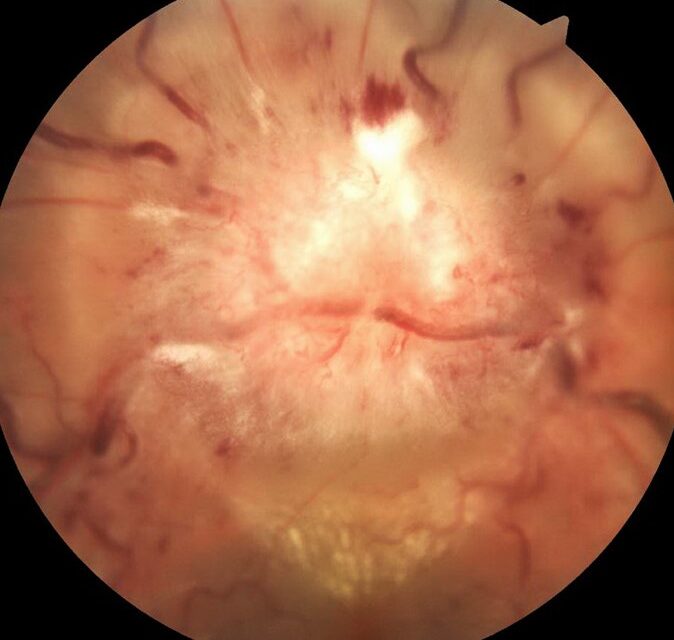
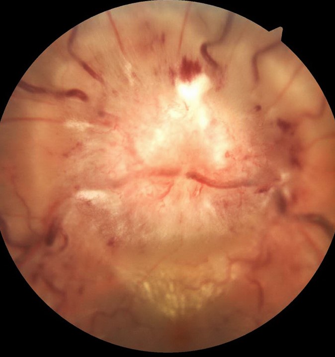
Fundus examination revealed optic disc swelling with a macular star (classic stellate maculopathy). Optical coherence tomography (OCT) demonstrated subretinal fluid and exudates in the macula, and fluorescein angiography (FA) confirmed disc leakage.
Serology for Bartonella henselae was positive, confirming the diagnosis of Bartonella henselae neuroretinitis.
The patient was treated with doxycycline and rifampin, leading to the gradual resolution of symptoms over three months.
Disease Entity
Bartonella henselae neuroretinitis is an infectious, inflammatory optic neuropathy characterized by unilateral optic disc swelling and macular exudates forming a star pattern.
It is one of the most common causes of infectious neuroretinitis and is strongly associated with cat scratch disease (CSD).
Fundus Explorer Pro
Photograph the retinal findings described here with the phone already in your pocket — 22 D optics and built-in illumination in one handheld unit.
From Choroida — the team behind this sitePathophysiology
Bartonella henselae is a gram-negative, intracellular bacterium transmitted through cat scratches, bites, or flea vectors.
The pathogen invades the vascular endothelium and immune cells, triggering an inflammatory response that affects the optic nerve and retina.
The hallmark findings—optic disc swelling and macular star—occur due to:
- Optic nerve inflammation leading to disc edema.
- Leakage of plasma lipoproteins into the Henle fiber layer, forming a macular star.
Epidemiology
- Age group: Primarily affects children and young adults.
- Transmission: Occurs via cat scratches, bites, or flea vectors.
- Prevalence: Common in areas with a high domestic cat population.

Clinical Features
Symptoms
- Sudden, painless vision loss (mild to moderate).
- Central or paracentral scotoma.
- Photopsia (flashes of light) in some cases.
- Mild systemic symptoms (low-grade fever, fatigue, lymphadenopathy).
Fundoscopic Findings
- Unilateral optic disc edema (hyperemic, swollen disc).
- Macular star (exudates forming a stellate pattern in the Henle fiber layer).
- Mild retinal vascular changes.
- Absence of vitritis (distinguishing it from infectious posterior uveitis).
Imaging Findings
- Optical Coherence Tomography (OCT):
- Thickened optic nerve head.
- Intraretinal hyperreflective deposits at the macula.
- Fluorescein Angiography (FA):
- Optic disc leakage without significant retinal vasculitis.
- Fundus Autofluorescence (FAF):
- Hyperautofluorescence at sites of exudation.
Differential Diagnosis
- Idiopathic Neuroretinitis: Lacks systemic Bartonella exposure history.
- Leber’s Hereditary Optic Neuropathy (LHON): Typically bilateral and progressive.
- Viral or Syphilitic Optic Neuritis: Often associated with vitritis or vasculitis.
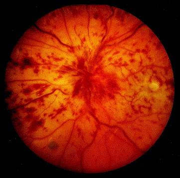
- Hypertensive Retinopathy: Features flame hemorrhages and cotton wool spots.
Diagnosis
- Clinical history (cat exposure, systemic symptoms).
- Serologic testing for Bartonella henselae IgM/IgG (enzyme-linked immunosorbent assay [ELISA] or polymerase chain reaction [PCR]).
- Ocular imaging (OCT, FA, FAF) to confirm neuroretinitis findings.

Management
Antimicrobial Therapy
- Doxycycline (100 mg BID) + Rifampin (300 mg BID) for 4-6 weeks.
- Alternative: Azithromycin, especially in children or pregnant patients.
Anti-inflammatory Treatment
- Oral corticosteroids (Prednisone 40-60 mg/day, tapered) if severe optic disc edema persists.
- Avoid early steroid use without antibiotics to prevent worsening infection.
Supportive Care
- Regular follow-up with OCT and visual field testing.
- Amsler grid home monitoring for early detection of progression.
Prognosis
- Generally favorable: Most patients recover vision within 2-6 months.
- Residual visual impairment: Some cases develop persistent optic disc pallor or mild scotomas.
Conclusion
Bartonella henselae neuroretinitis is an infectious optic neuropathy presenting with sudden vision loss, optic disc swelling, and macular star formation. Diagnosis is based on clinical history, serologic testing, and imaging. Early antibiotic therapy leads to excellent recovery, making timely recognition essential.


Document what you see
Two smartphone imaging tools built for everyday clinic use — one for the slit lamp, one for the fundus.
From Choroida — the team behind this siteReferences
- Solley WA, Martin DF, Newman NJ, et al. “Cat scratch disease: posterior segment manifestations.” Ophthalmology. 1999;106(8):1546-1553.
- Reed JB, Scales DK, Wong MT, et al. “Neuroretinitis: A review of the literature and new observations.” Surv Ophthalmol. 1998;43(2):155-180.
- Mazur-Melewska K, Mania A, Kemnitz P, et al. “Cat scratch disease: A wide spectrum of ocular manifestations.” Eur J Pediatr. 2015;174(9):1193-1200.
- Ormerod LD, Dailey JP. “Ocular manifestations of cat-scratch disease.” Curr Opin Ophthalmol. 1999;10(3):209-216.
Test yourself
A few questions straight from this article.
-
How is Bartonella henselae neuroretinitis best defined?
It is an infectious inflammatory optic neuropathy with unilateral optic disc swelling and macular exudates arranged in a star, and the commonest infectious neuroretinitis. -
Which systemic illness is Bartonella henselae neuroretinitis most strongly associated with?
Bartonella henselae neuroretinitis is strongly linked to cat scratch disease, acquired through cat scratches, bites or flea vectors. -
What type of organism is Bartonella henselae?
Bartonella henselae is a gram-negative intracellular bacterium that invades vascular endothelium and immune cells, provoking inflammation of the optic nerve and retina. -
The macular star of Bartonella neuroretinitis forms from lipoprotein leakage into which layer?
Plasma lipoproteins leak into the Henle fibre layer, where the radial arrangement of fibres produces the classic stellate macular exudate pattern. -
Which age group is chiefly affected by Bartonella henselae neuroretinitis?
The condition primarily affects children and young adults and is commoner in regions with a large domestic cat population. -
Which fundus feature helps separate Bartonella neuroretinitis from infectious posterior uveitis?
The absence of vitritis distinguishes Bartonella neuroretinitis from infectious posterior uveitis, even though the disc is hyperaemic and swollen. -
What does fluorescein angiography typically demonstrate in Bartonella henselae neuroretinitis?
FA shows optic disc leakage without significant retinal vasculitis, while OCT reveals a thickened optic nerve head and intraretinal hyperreflective macular deposits. -
Which antibiotic regimen is first-line for Bartonella henselae neuroretinitis in adults?
Doxycycline 100 mg twice daily with rifampin 300 mg twice daily for four to six weeks is standard, with azithromycin used in children and pregnancy. -
How should corticosteroids be used in Bartonella henselae neuroretinitis?
Oral prednisone 40-60 mg daily with a taper may be added if severe disc oedema persists, but early steroids without antibiotic cover risk worsening the infection. -
What is the usual visual prognosis of Bartonella henselae neuroretinitis?
The prognosis is generally favourable, with most patients recovering vision within two to six months, though some retain optic disc pallor or mild scotomas.
