
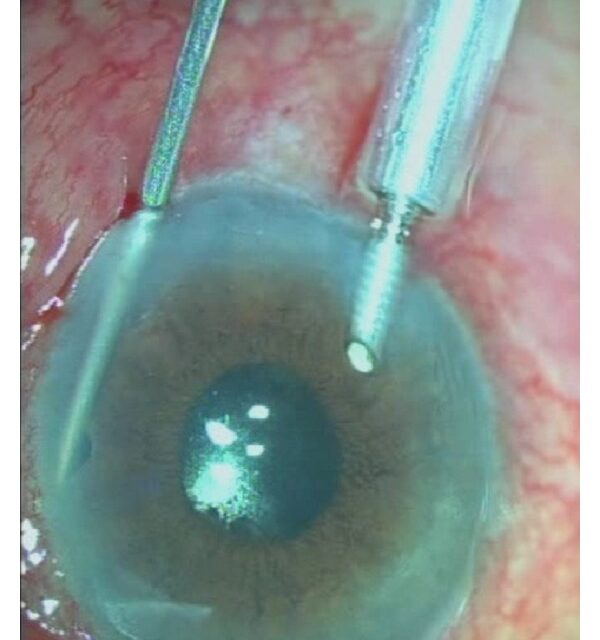
Malignant glaucoma, also called aqueous misdirection syndrome or ciliary block glaucoma, is a rare but genuinely dangerous postoperative complication in which aqueous humor is diverted posteriorly into and behind the vitreous instead of flowing forward into the anterior chamber.
The name is a historical artifact, since the condition has nothing to do with malignancy in the oncologic sense, but the label has stuck because of how aggressively it can progress and how easily it can be mistaken for a much more common, very differently treated problem.
Confusing malignant glaucoma with pupillary block angle closure is the single most important pitfall to avoid, since the standard treatment for pupillary block actively worsens malignant glaucoma.
The condition is uncommon overall, but it disproportionately affects eyes that already carried some predisposing anatomic risk before their original surgery, which is part of why a careful preoperative assessment of anterior segment crowding has some value in anticipating it.
Mechanism
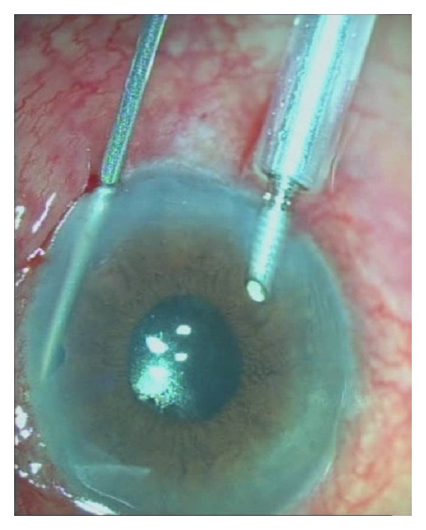
In malignant glaucoma, a misdirection of aqueous flow occurs at the level of the ciliary body, vitreous face, and lens-iris diaphragm, causing aqueous to accumulate posteriorly rather than passing forward through the pupil into the anterior chamber in the normal direction.
This posterior fluid accumulation pushes the entire lens-iris diaphragm forward, shallowing or flattening the anterior chamber uniformly, including centrally, which is the key finding that separates it from pupillary block.
The exact anatomic trigger is thought to involve anterior rotation of the ciliary processes crowding the space around the lens equator and anterior vitreous face, though the precise mechanism in any individual case can be difficult to pin down with certainty.
Risk Factors and Setting
- Recent intraocular surgery, most classically glaucoma filtering surgery, though it can also follow cataract surgery or other intraocular procedures
- A history of angle-closure glaucoma or a predisposing anatomic configuration, including a short axial length and crowded anterior segment
- Prior malignant glaucoma in the fellow eye, which substantially raises the risk of the same process occurring in the second eye during or after its own surgery
- Use of miotic medications, which can precipitate or worsen the condition by further crowding an already anatomically predisposed anterior segment
All-fit Slit-Lamp Adapter
Record and share exactly what you see at the slit lamp. One adapter fits any slit lamp or surgical microscope — and any smartphone.
From Choroida — the team behind this siteDistinguishing It From Pupillary Block
Both conditions present with a markedly elevated intraocular pressure and a shallow anterior chamber, which is exactly why the distinction is so easy to miss without deliberately looking for it.
In pupillary block, the peripheral anterior chamber is shallow while the central chamber, where the lens sits directly behind an open pupil, can be relatively less affected, and a peripheral iridotomy resolves the block by giving aqueous an alternate forward path.
In malignant glaucoma, the entire anterior chamber, both centrally and peripherally, is uniformly shallow or flat, and a patent iridotomy, whether pre-existing or newly performed, does not relieve the pressure, since the problem is not a block at the pupil at all.
A shallow chamber that persists or worsens despite a confirmed patent iridotomy is the clinical clue that should immediately raise suspicion for malignant glaucoma rather than an incompletely treated pupillary block.
Management
Cycloplegic agents, particularly atropine, are first-line treatment, working by tightening zonular tension and pulling the lens-iris diaphragm posteriorly, which is the opposite of what miotics would do and is precisely why the two are managed so differently.
Aqueous suppressants reduce production of the fluid that is accumulating in the wrong compartment, providing symptomatic pressure control while the cycloplegic treatment works to correct the underlying anatomic misdirection.
Medical therapy resolves a meaningful proportion of cases, but eyes that do not respond require surgical intervention, most often vitrectomy with disruption of the anterior hyaloid face to break the cycle of posterior fluid trapping directly.
In a phakic eye that fails medical treatment, lens extraction combined with vitrectomy and anterior hyaloidotomy is often needed, since the lens itself contributes to the anatomic crowding driving the condition.
Why Prompt Recognition Matters
Malignant glaucoma is a genuine emergency, since sustained, severely elevated intraocular pressure threatens the optic nerve in the same way any other severe pressure spike does, on top of the risk of corneal decompensation from a persistently shallow chamber.
Delayed recognition, particularly when the condition is mistakenly treated as pupillary block with miotics, can actively worsen the anatomic crowding and delay the correct treatment while vision continues to be at risk.
Given the elevated risk to a fellow eye undergoing similar surgery later, that risk should be explicitly documented and discussed with the patient before any future intraocular procedure on the other side.


Document what you see
Two smartphone imaging tools built for everyday clinic use — one for the slit lamp, one for the fundus.
From Choroida — the team behind this siteReferences
- Ruben S, Tsai J, Hitchings RA. Malignant glaucoma and its management. British Journal of Ophthalmology.
- American Academy of Ophthalmology. Basic and Clinical Science Course, Section 10: Glaucoma.
- Debrouwere V, Stalmans P, Van Calster J, et al. Outcomes of different management options for malignant glaucoma: a retrospective study. Graefe’s Archive for Clinical and Experimental Ophthalmology.
