
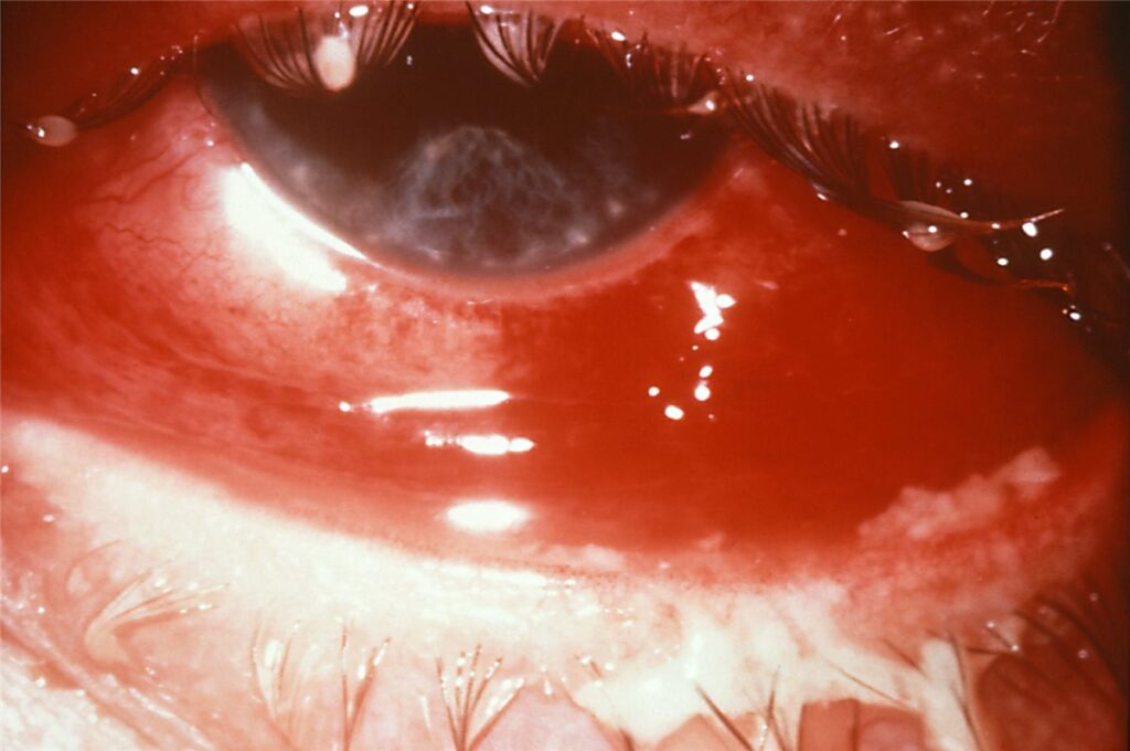
Adult gonococcal conjunctivitis is a hyperacute bacterial conjunctivitis caused by Neisseria gonorrhoeae, almost always acquired through autoinoculation from genital secretions rather than direct droplet spread, and it is one of the few forms of conjunctivitis that can perforate a cornea within days if it is dismissed as routine.
The pace of this disease is what separates it from the conjunctivitis seen in clinic every day. What starts as redness and discharge can progress to corneal ulceration and perforation over 24 to 48 hours, which is why recognising the hyperacute pattern matters more than remembering the organism’s name.
Why This Organism Behaves Differently
Neisseria gonorrhoeae is one of the few bacteria capable of penetrating an intact corneal epithelium, unlike the great majority of organisms causing bacterial conjunctivitis, which require a break in the epithelial barrier to invade the cornea.
This direct invasive capacity, combined with an intense neutrophilic inflammatory response, is what accounts for the speed and severity of corneal involvement, and it is the central reason this organism is managed so differently from routine bacterial conjunctivitis.
Clinical Presentation
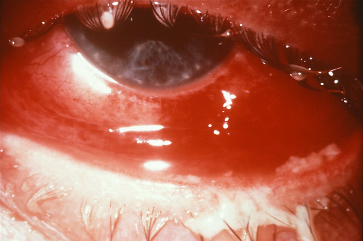
Onset is rapid, typically within 12 to 24 hours of exposure, with copious, thick purulent discharge that reaccumulates within minutes of being wiped away, a feature that is genuinely distinctive once seen and a useful clue at the bedside.
Marked conjunctival injection, chemosis, and lid swelling, sometimes severe enough to make the eye difficult to open, accompany the discharge. Preauricular lymphadenopathy is common and, when present alongside hyperacute purulent discharge, further supports the diagnosis over more routine bacterial causes.
Pain and photophobia beyond what would be expected for simple conjunctivitis suggest corneal involvement and should prompt urgent slit-lamp assessment specifically for epithelial defect, infiltrate, or thinning.
All-fit Slit-Lamp Adapter
Record and share exactly what you see at the slit lamp. One adapter fits any slit lamp or surgical microscope — and any smartphone.
From Choroida — the team behind this siteExamination Findings
- Copious, thick, yellow-green purulent discharge under visible pressure, reaccumulating rapidly after cleaning
- Marked papillary conjunctival reaction and chemosis
- Corneal epithelial defect, infiltrate, or, in severe cases, frank ulceration, sometimes progressing to perforation if untreated
- Preauricular lymphadenopathy
- Signs of concurrent genital infection should be asked about directly, since the source of ocular infection is almost always autoinoculation from genital secretions
Diagnostic Evaluation
Conjunctival swab for Gram stain, which classically shows Gram-negative intracellular diplococci within neutrophils, allows a rapid presumptive diagnosis that can guide immediate treatment while culture results are pending.
Culture on selective media, such as chocolate or Thayer-Martin agar, confirms the diagnosis and allows antimicrobial susceptibility testing, which is increasingly relevant given rising rates of antimicrobial resistance in Neisseria gonorrhoeae.
Nucleic acid amplification testing is highly sensitive and increasingly used alongside culture. Testing for concurrent chlamydial infection is appropriate given frequent coinfection, and broader sexually transmitted infection screening, including HIV and syphilis, should be offered as part of the same visit.
Differential Diagnosis
- Other bacterial conjunctivitis, generally with a slower onset and less copious discharge, without the same corneal perforation risk
- Viral conjunctivitis, typically watery rather than purulent discharge, and without the intracellular diplococci seen on Gram stain
- Chlamydial conjunctivitis, more indolent in onset, though frequently coexisting with gonococcal infection and needing to be tested for regardless
- Allergic conjunctivitis, itchy rather than painful, with a clear rather than purulent discharge
The speed of onset and the volume of discharge are what most reliably separate this from other causes of a red eye, and either finding alone in a sexually active adult should prompt urgent same-day assessment rather than routine review.
Management
Systemic Antibiotics
Systemic therapy is essential; topical treatment alone is inadequate for gonococcal conjunctivitis given the organism’s capacity for corneal invasion. A single dose of an appropriate third-generation cephalosporin, typically intramuscular or intravenous ceftriaxone, is standard first-line treatment, with the specific regimen guided by current local antimicrobial resistance patterns and guidelines.
Local Measures
Frequent saline irrigation of the conjunctival sac helps clear purulent discharge and reduce the bacterial load in contact with the ocular surface, and topical antibiotics are typically used as an adjunct to systemic treatment.
Treating Coinfection and Partners
Empirical treatment for chlamydial coinfection is standard practice given how frequently the two occur together. Sexual partners require assessment and treatment, and referral to sexual health services is an appropriate part of management, not an optional extra.
Monitoring for Corneal Involvement
Close follow-up with repeat slit-lamp examination over the following days is warranted to confirm resolution and catch any progression of corneal involvement early, particularly in patients who present with any degree of corneal findings at the initial visit.
Prognosis
With prompt systemic treatment, adult gonococcal conjunctivitis generally resolves without long-term visual sequelae.
Delayed diagnosis or treatment risks progression to corneal ulceration and, in severe cases, perforation, which can result in permanent scarring or loss of the eye, an outcome that is now largely preventable with timely recognition and systemic antibiotic therapy.
Because this condition can move from mild redness to a corneal emergency within a day or two, the practical lesson is that any adult with hyperacute, copious purulent discharge deserves same-day assessment and, where the diagnosis is suspected, treatment started before laboratory confirmation returns.


Document what you see
Two smartphone imaging tools built for everyday clinic use — one for the slit lamp, one for the fundus.
From Choroida — the team behind this siteReferences
- Workowski KA, Bachmann LH, Chan PA, et al. Sexually Transmitted Infections Treatment Guidelines. MMWR Recommendations and Reports. 2021.
- Ullman S, Roussel TJ, Forster RK. Gonococcal keratoconjunctivitis. Survey of Ophthalmology. 1987.
- World Health Organization. WHO guidelines for the treatment of Neisseria gonorrhoeae.
- Gonococcal Conjunctivitis. EyeWiki, American Academy of Ophthalmology.
- Gonococcal Conjunctivitis. StatPearls, NCBI Bookshelf.
Test yourself
A few questions straight from this article.
-
How is adult gonococcal conjunctivitis almost always acquired?
The ocular infection is almost always autoinoculated from genital secretions rather than spread by droplets, which is why concurrent genital infection should be asked about directly. -
Which property of Neisseria gonorrhoeae explains the rapid corneal involvement it causes?
Unlike most conjunctivitis organisms, which need a break in the epithelial barrier, Neisseria gonorrhoeae can invade through intact epithelium, and the intense neutrophilic response compounds the damage. -
How soon after exposure does adult gonococcal conjunctivitis typically begin?
Onset is typically within 12 to 24 hours of exposure, and corneal ulceration can follow over the next 24 to 48 hours if treatment is delayed. -
Which discharge characteristic is distinctive of adult gonococcal conjunctivitis?
The thick purulent discharge is under visible pressure and refills within minutes of being wiped away, a genuinely distinctive bedside clue. -
What does conjunctival Gram stain classically show in adult gonococcal conjunctivitis?
Gram stain classically shows Gram-negative intracellular diplococci within neutrophils, allowing a rapid presumptive diagnosis while culture is pending. -
Which selective culture medium confirms gonococcal conjunctivitis and allows susceptibility testing?
Culture on selective media such as chocolate or Thayer-Martin agar confirms the diagnosis and gives susceptibility data, increasingly relevant with rising gonococcal resistance. -
Which feature points to viral rather than gonococcal conjunctivitis in an adult?
Viral conjunctivitis gives watery rather than copious purulent discharge and lacks the intracellular diplococci seen on Gram stain in gonococcal disease. -
What is standard first-line treatment for adult gonococcal conjunctivitis?
Systemic therapy is essential because topical treatment alone cannot control an organism capable of corneal invasion; a single dose of intramuscular or intravenous ceftriaxone is standard, guided by local resistance patterns. -
What should be treated empirically alongside adult gonococcal conjunctivitis?
Chlamydial coinfection is frequent enough that empirical cover is standard practice, alongside partner assessment and broader sexual health screening. -
What is the consequence of delayed treatment in adult gonococcal conjunctivitis?
Delay risks progression to corneal ulceration and perforation, with permanent scarring or loss of the eye, an outcome largely preventable with prompt systemic antibiotics.

Writes and reviews the clinical articles on Educate. Every post is checked against peer-reviewed sources before it goes live.


