
Radial keratotomy, or RK, was the first widely adopted refractive surgery for myopia, using a pattern of deep radial corneal incisions to flatten the central cornea and reduce nearsightedness.
It was largely abandoned once excimer laser techniques like PRK and LASIK arrived, offering more predictable, more stable outcomes with far fewer long-term surprises.
RK’s real relevance today lies less in the procedure itself, which almost nobody still performs, than in the population of patients who had it decades ago and are now presenting for cataract surgery with a cornea that behaves very differently from an untouched one.
RK was performed on a very large number of patients during its peak popularity in the 1980s and early 1990s, and enough time has now passed that a meaningful share of that original patient population has reached typical cataract age.
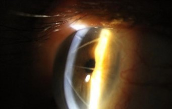
Technique
The surgeon made a series of deep, radially oriented incisions in the peripheral and mid-peripheral cornea, sparing a central optical zone, using a calibrated diamond blade to control incision depth.
As the peripheral cornea weakened along these incision lines, it bulged outward under normal intraocular pressure, causing the central cornea to flatten and reducing the eye’s overall refractive power.
The number, depth, and length of the incisions were varied based on the amount of myopia being corrected, with more aggressive incision patterns used for higher degrees of myopia.
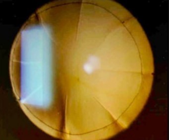
A typical pattern used four to eight incisions, though the exact number and configuration varied considerably by surgeon and by the specific nomogram in use at the time.
Why It Fell Out of Favor
Refractive outcomes were considerably less predictable than what excimer laser techniques later achieved, with results varying meaningfully between surgeons and even between eyes of the same patient.
A progressive hyperopic shift over subsequent years was a well-documented long-term problem, as the weakened peripheral cornea continued to remodel slowly over time, gradually overcorrecting the original myopia.
Diurnal fluctuation in vision, with refraction measurably different in the morning compared to later in the day, was another recognized complication tied to the cornea’s altered biomechanics.
Corneal weakening from the incisions also raised concerns about structural stability under any future stress, a concern that has become directly relevant now that many RK patients are reaching the age for cataract surgery.
By the time excimer laser platforms became widely available and refined, RK’s comparatively unpredictable results made it difficult to justify over the newer technology for most surgeons and patients alike.
All-fit Slit-Lamp Adapter
Record and share exactly what you see at the slit lamp. One adapter fits any slit lamp or surgical microscope — and any smartphone.
From Choroida — the team behind this siteRelevance to Modern Cataract Surgery
IOL power calculation is genuinely more difficult in a post-RK eye, since standard formulas assume a cornea with a normal, unaltered relationship between anterior and posterior curvature, an assumption RK incisions violate.
Post-refractive-surgery IOL calculation methods, using either historical preoperative data when available or specialized formulas designed for eyes with prior corneal refractive surgery, are needed to reduce the risk of a significant postoperative refractive surprise.
- Standard keratometry tends to underestimate the true corneal power in a post-RK eye, which without correction leads to a hyperopic surprise after cataract surgery
- Historical data from before the RK procedure, including preoperative keratometry and refraction, meaningfully improves calculation accuracy when the patient or their prior records can supply it
- Topography-guided and post-refractive-specific IOL formulas have been developed specifically to address the altered corneal shape in these eyes
Intraoperative Considerations
A weakened, incised cornea from RK is also at some risk of incision opening or wound dehiscence during cataract surgery itself, from the mechanical stress of the phacoemulsification procedure and fluctuations in intraocular pressure during the case.
Surgeons operating on a post-RK eye typically take extra care with wound construction and pressure management, and counsel the patient in advance that both the refractive outcome and the surgical course itself carry more uncertainty than in an eye without a prior refractive procedure.
Patients should be counseled clearly before surgery that a post-RK eye carries a meaningfully higher chance of needing a refractive touch-up or an IOL exchange afterward compared to an eye with normal, unoperated corneal anatomy.
Many of these patients remember their original RK surgery as having been genuinely difficult to predict, and setting expectations for cataract surgery that acknowledges that same history tends to go over better than presenting the new surgery as entirely routine.


Document what you see
Two smartphone imaging tools built for everyday clinic use — one for the slit lamp, one for the fundus.
From Choroida — the team behind this siteReferences
- Waring GO 3rd, Lynn MJ, McDonnell PJ. Results of the prospective evaluation of radial keratotomy (PERK) study 10 years after surgery. Archives of Ophthalmology.
- American Academy of Ophthalmology. Basic and Clinical Science Course, Section 13: Refractive Surgery.
- Wang L, Booth MA, Koch DD. Comparison of intraocular lens power calculation methods in eyes that have undergone LASIK. Ophthalmology.
