
Hemi-central retinal vein occlusion is an occlusion confined to one of the two trunks of the central retinal vein, producing a pattern of hemorrhage and edema that sits anatomically between a branch retinal vein occlusion and a full central retinal vein occlusion.
It exists because, in a meaningful minority of eyes, the central retinal vein bifurcates into two separate trunks before it exits through the lamina cribrosa rather than remaining a single trunk, and either half can occlude independently of the other.
Recognizing this variant matters because its prognosis and behavior do not map cleanly onto either of the two occlusions it superficially resembles.
It is an uncommon presentation overall, and many general ophthalmologists go through training without ever having the anatomic variant specifically pointed out to them, which makes misclassification as an atypical branch or central occlusion fairly common.
Anatomic Basis
In eyes where the anatomic variant is present, the central retinal vein is effectively duplicated proximal to the lamina cribrosa, with each trunk draining roughly one half of the retina, typically split along a superior-inferior axis.
An occlusion of one trunk produces hemorrhage and venous congestion confined to that half of the retina, while the other half, drained by the unaffected trunk, remains largely unaffected.
This is mechanically distinct from a branch retinal vein occlusion, where the occlusion occurs more peripherally at an arteriovenous crossing, and from a full central retinal vein occlusion, where both trunks (or a single undivided trunk) are occluded together.
Not every eye has this dual-trunk anatomy, which is why hemi-central retinal vein occlusion is inherently less common than either of the two occlusions that flank it in severity.
Clinical Presentation
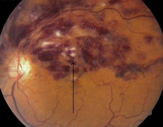
- Hemorrhages, venous dilation, and cotton wool spots confined to one hemisphere of the retina, typically split along the horizontal raphe
- Macular edema when the affected hemisphere includes the papillomacular bundle or a significant portion of the central macula
- Vision loss proportional to macular involvement, ranging from mild if the fovea is spared to significant if the occlusion involves the hemisphere containing central fixation
- A disc appearance that can show hemorrhage or swelling limited to the corresponding half, rather than the more diffuse disc changes typical of a full central occlusion
Fundus Explorer Pro
Photograph the retinal findings described here with the phone already in your pocket — 22 D optics and built-in illumination in one handheld unit.
From Choroida — the team behind this siteDistinguishing It From BRVO and CRVO
The extent of retina involved is the key distinguishing feature: a branch occlusion affects a wedge distal to a specific arteriovenous crossing, a hemi-central occlusion affects roughly half the retina along the horizontal meridian, and a full central occlusion affects the entire retina.
Fluorescein angiography helps confirm the distribution of nonperfusion and delayed filling, which typically respects the horizontal midline in a hemi-central occlusion in a way that neither a branch nor a full central occlusion does.
This distinction is not just academic, since the extent of retinal involvement correlates with the risk of both macular edema and neovascular complications, and management decisions are made hemisphere by hemisphere rather than assuming the whole retina behaves as one unit.
Ischemic Risk and Complications
Hemi-central retinal vein occlusion carries an intermediate risk of neovascular complications compared with branch and full central occlusions, broadly tracking with how much retinal capillary nonperfusion is present on angiography in the affected hemisphere.
Neovascularization of the iris or angle, when it occurs, tends to behave similarly to the ischemic form of full central retinal vein occlusion and warrants the same vigilance and treatment threshold.
Regular monitoring for neovascularization, including gonioscopy at follow-up visits, is warranted in eyes with significant nonperfusion on angiography, following the same general framework used for full central retinal vein occlusion.
Because only half the retina is at risk rather than the whole posterior pole, some eyes with a hemi-central occlusion do better than their angiographic nonperfusion count might suggest if extrapolated directly from full central retinal vein occlusion data, which is a distinction worth remembering when counseling patients on prognosis.
Management
Macular edema is treated the same way it is in branch and central retinal vein occlusion, primarily with intravitreal anti-VEGF therapy, with intravitreal corticosteroids as an alternative or adjunct in selected cases.
Panretinal photocoagulation is reserved for eyes that develop neovascularization or show extensive nonperfusion on angiography, mirroring the threshold used in ischemic central retinal vein occlusion.
Systemic risk factor assessment, including blood pressure, glucose, and lipid evaluation, is appropriate for any retinal vein occlusion, since the underlying vascular risk factors are shared across all three anatomic subtypes.
Follow-up intervals generally track the extent of macular involvement and the degree of nonperfusion, with more ischemic-appearing hemi-central occlusions followed at least as closely as a comparable ischemic central retinal vein occlusion would be.


Document what you see
Two smartphone imaging tools built for everyday clinic use — one for the slit lamp, one for the fundus.
From Choroida — the team behind this siteReferences
- Hayreh SS. Hemi-central retinal vein occlusion: pathogenesis, clinical features, and natural history. Archives of Ophthalmology.
- American Academy of Ophthalmology. Basic and Clinical Science Course, Section 12: Retina and Vitreous.
- The Central Vein Occlusion Study Group. Natural history and clinical management of central retinal vein occlusion. Archives of Ophthalmology.
Test yourself
A few questions straight from this article.
-
Hemi-central retinal vein occlusion involves occlusion of which structure?
The occlusion is confined to one of the two trunks of the central retinal vein, giving a pattern of haemorrhage and oedema that sits between branch and full central occlusion. -
Which anatomic variant allows hemi-central retinal vein occlusion to occur?
In a minority of eyes the central retinal vein bifurcates into two trunks before exiting through the lamina cribrosa, each draining about half the retina, so either half can occlude alone. -
What disc appearance is typical of hemi-central retinal vein occlusion?
The disc may show haemorrhage or swelling confined to the half matching the occluded trunk, rather than the diffuse disc changes typical of a full central retinal vein occlusion. -
How does hemi-central retinal vein occlusion differ anatomically from a branch retinal vein occlusion?
A branch occlusion happens more peripherally at an arteriovenous crossing and affects a wedge distal to it, whereas a hemi-central occlusion blocks one venous trunk at the disc and affects half the retina. -
Which angiographic pattern supports a diagnosis of hemi-central retinal vein occlusion?
Fluorescein angiography shows nonperfusion and delayed filling that respect the horizontal midline, a distribution neither a branch nor a full central occlusion produces. -
What is the risk of neovascular complications in hemi-central retinal vein occlusion?
The neovascular risk is intermediate between branch and full central occlusion and broadly tracks the amount of capillary nonperfusion in the affected hemisphere. -
In hemi-central retinal vein occlusion with significant nonperfusion, which examination is repeated at follow-up?
Eyes with significant nonperfusion need regular monitoring for neovascularisation, including gonioscopy at follow-up, using the same framework applied to full central retinal vein occlusion. -
How is macular oedema from hemi-central retinal vein occlusion treated?
Macular oedema is managed exactly as in branch and central occlusion, primarily with intravitreal anti-VEGF therapy and intravitreal corticosteroid as an alternative or adjunct in selected eyes. -
When is panretinal photocoagulation used in hemi-central retinal vein occlusion?
Panretinal photocoagulation is reserved for eyes that develop neovascularisation or show extensive nonperfusion, mirroring the threshold used in ischemic central retinal vein occlusion. -
Which systemic assessment is appropriate after any retinal vein occlusion?
The vascular risk factors are shared across all three anatomic subtypes, so blood pressure, glucose and lipid assessment is appropriate for any retinal vein occlusion.

Writes and reviews the clinical articles on Educate. Every post is checked against peer-reviewed sources before it goes live.


